. 2015 May;27(5):1389-408.
doi: 10.1105/tpc.15.00158. Epub 2015 May 5.

Autophagic recycling plays a central role in maize nitrogen remobilization

Affiliations

Autophagic recycling plays a central role in maize nitrogen remobilization

Faqiang Li et al. Plant Cell. 2015 May.

Abstract

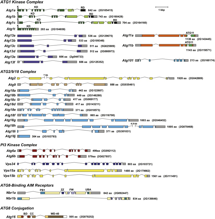
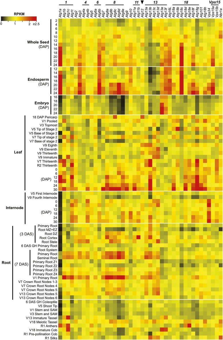
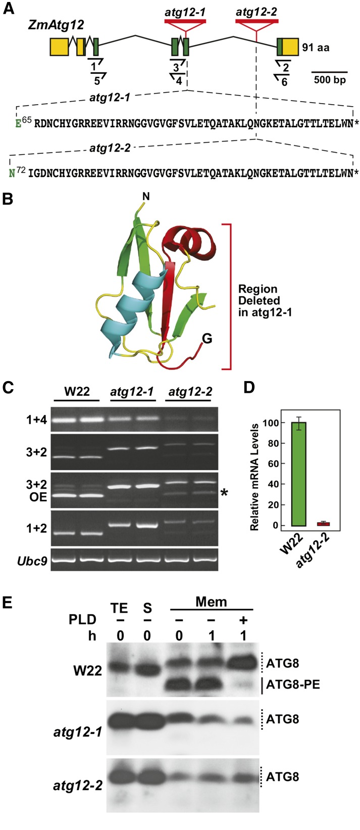
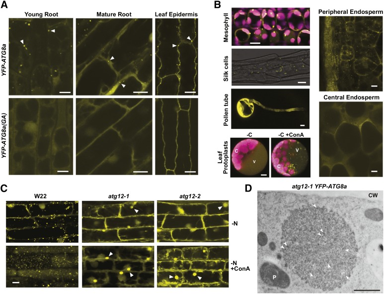
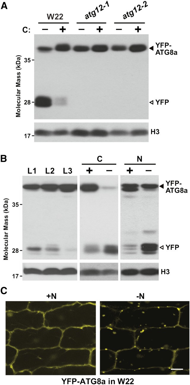
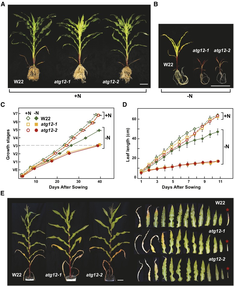
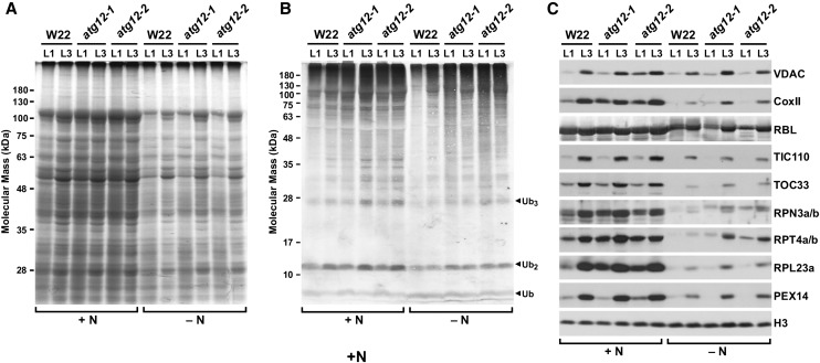
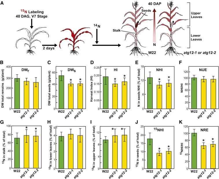
Autophagy is a primary route for nutrient recycling in plants by which superfluous or damaged cytoplasmic material and organelles are encapsulated and delivered to the vacuole for breakdown. Central to autophagy is a conjugation pathway that attaches AUTOPHAGY-RELATED8 (ATG8) to phosphatidylethanolamine, which then coats emerging autophagic membranes and helps with cargo recruitment, vesicle enclosure, and subsequent vesicle docking with the tonoplast. A key component in ATG8 function is ATG12, which promotes lipidation upon its attachment to ATG5. Here, we fully defined the maize (Zea mays) ATG system transcriptionally and characterized it genetically through atg12 mutants that block ATG8 modification. atg12 plants have compromised autophagic transport as determined by localization of a YFP-ATG8 reporter and its vacuolar cleavage during nitrogen or fixed-carbon starvation. Phenotypic analyses showed that atg12 plants are phenotypically normal and fertile when grown under nutrient-rich conditions. However, when nitrogen-starved, seedling growth is severely arrested, and as the plants mature, they show enhanced leaf senescence and stunted ear development. Nitrogen partitioning studies revealed that remobilization is impaired in atg12 plants, which significantly decreases seed yield and nitrogen-harvest index. Together, our studies demonstrate that autophagy, while nonessential, becomes critical during nitrogen stress and severely impacts maize productivity under suboptimal field conditions.


Figures

Figure 1.
Figure 1.
Annotation of Genes Encoding Upstream Components of the Maize ATG8-Mediated Autophagic System. Included are genes encoding components from the ATG1 kinase complex, the ATG9/2/18 complex, the PI3 kinase complex, the ubiquitin receptor NBR1 that helps recognize ubiquitylated substrates during selective autophagy, and ATG16 that associates with the ATG12-ATG5 conjugate and promotes formation of the ATG8-PE adduct. The amino acid sequence length and maize genome GRMZM accession number of each protein/gene are on the right. Colored and gray boxes denote coding regions and untranslated regions, respectively. Lines indicate introns. The conserved lysine (K) within the ATP hydrolysis site in the ATG1 kinase family is indicated by arrowheads. The kinase domain (KD) and regulatory domain (RD) in ATG1, the ATG11 domain in ATG11, and the Phox and Bem1p (PB1), ZZ-type zinc finger (ZZ), four tryptophan (FW), and ubiquitin-associated (UBA) domains in NBR1 are shown. See Supplemental Table 1 for more information on each gene.
Figure 2.
Figure 2.
Developmental and Tissue-Specific Expression Profiles of Maize Atg Genes. RNA-seq experiments representing 80 developmentally or anatomically distinct maize samples were analyzed for autophagy-related genes based on reads per kilobase per one million reads (RPKM). Gene families encoding individual ATG factors are highlighted on the left. The arrowhead locates transcript values from the single maize Atg12 locus. The color indicates the degree of fold change: red, high; black, low. Vegetative (V1 to 18) and reproductive (R1 and 2) growth stages were defined based on the Maize Field Guide published by Iowa State University Extension (Abendroth et al., 2011). Dissected primary root: Z1, zone 1 (first centimeter of root tip); Z2, zone 2 (from end of Z1 to the point of root hair/lateral root initiation); Z3, zone 3 (lower half of differentiation zone); Z4, zone 4 (upper half of differentiation zone). DZ, differentiation zone; EZ, elongation zone; MZ, meristematic zone; SAM, shoot apical meristem. See Supplemental Table 2 for full descriptions of the tissues analyzed.
Figure 3.
Figure 3.
Genetic Description of Maize atg12 Mutants (A) Gene diagram of the maize Atg12 locus showing its exon/intron organization and the positions of atg12-1 and atg12-2 UniformMu insertion mutations. Lines represent introns and the green and yellow boxes indicate coding and untranslated regions, respectively. Half arrows underneath locate positions of the primers used for RT-PCR in (C) and quantitative RT-PCR in (D). Amino acids in black identify the codons appended after Glu-65 in atg12-1 and Asn-72 in atg12-2, respectively. Asterisks indicate stop codons. (B) Three-dimensional ribbon diagram of Arabidopsis ATG12 (PDB 1WZ3). The polypeptide region deleted by the atg12-1 insertion is colored in red. (C) RT-PCR analysis demonstrating the accumulation of longer, Mu element-containing transcripts in homozygous atg12-1 and atg12-2 seedlings. Wild-type W22 was included as a control. Positions of the primers used are shown in (A). OE, overexposure of the ethidium bromide-stained gel for the PCR products revealed that a wild type-like transcript (asterisk) accumulates in atg12-2 plants. RT-PCR of the UBC9 locus was used to verify the analysis of equal amounts of RNA. Two independent experiments gave similar results. (D) Quantitative real-time RT-PCR analysis of wild type-like atg12 transcripts in homozygous atg12-2 plants. The locations of primers 5 and 6 used for the PCR are shown in (A). Each bar represents mean (±sd) of three biological replicates. (E) ATG8 lipidation is blocked in the atg12 mutants. Seedlings were grown hydroponically on high-N liquid medium for 10 d and then exposed to low-N medium for 2 d before sampling. Total protein extracts (TE) prepared from root tissues were separated into soluble (S) and membrane fractions (Mem) by centrifugation, and the membrane fractions were solubilized with Triton X-100 and incubated with or without PLD for 1 h. Samples were subjected to SDS-PAGE in the presence of 6 M urea and immunoblotted with antibodies against Arabidopsis ATG8a. Solid and dashed lines locate ATG8-PE and free ATG8, respectively.
Figure 4.
Figure 4.
Autophagic Vesicles Detected Using the YFP-ATG8a Reporter Are Absent in atg12 Mutants. (A) and (B) Confocal fluorescence microscopy of maize B73 tissues expressing YFP-ATG8a or the YFP-ATG8a(GA) mutant. In (A), seedlings were grown hydroponically for 12 d in high-N liquid medium for imaging root cells or on soil for 14 d for imaging leaf epidermal cells. Possible autophagosomes observed in YFP-Atg8a plants are located by the arrowheads. In (B), images of mesophyll cells were captured from leaves of 2-week-old soil-grown seedlings. Chlorophyll fluorescence is shown in magenta. Silk, peripheral, and central endosperm tissues were collected 18 DAP from greenhouse-grown plants. For pollen tubes, fresh pollen grains were dusted on solid pollen germination medium and allowed to germinate for 4 h at room temperature before microscopy. For maize leaf protoplasts, the protoplasts from 2-week-old, soil-grown seedlings expressing YFP-ATG8a were incubated in Suc-free medium supplemented with 1 μM ConA for 16 h before microscopy. C, chloroplast; V, vacuole. Bars = 10 μm. (C) Maize atg12 mutants fail to accumulate autophagic bodies. Wild-type W22, atg12-1, and atg12-2 seedlings expressing YFP-ATG8a were grown hydroponically in high-N liquid medium for 10 d, transferred to low-N liquid medium for 32 h, and then treated for 16 h with 1 μM ConA or an equal volume of DMSO. Root cells were imaged by confocal fluorescence microscopy. The large amorphous cytoplasmic structures containing YFP-ATG8a that accumulate in atg12 plants are indicated by arrowheads. Bar = 10 μm. (D) Electron microscopy image of an atg12-1 root cell pretreated with ConA showing a YFP-ATG8a aggregate. Root tips were fixed, sliced into thin sections, and immunogold-labeled with anti-GFP antibodies (arrowheads). CW, cell wall; P, plastid. See Supplemental Figure 6A for additional images. Bar = 1 μm.
Figure 5.
Figure 5.
Autophagy Is Accelerated by Nitrogen or Fixed-Carbon Starvation in Maize. (A) atg12 mutants block the release of free YFP from YFP-ATG8a. Wild-type W22, atg12-1, and atg12-2 seedlings expressing YFP-ATG8a were grown on soil for 2 weeks and the second leaf blade (L2) was either kept in the light (+) or subjected to fixed-carbon (C) starvation by covering a section with aluminum foil for 2 d (−). Total protein extracted from a mix of three to six leaves was subjected to SDS-PAGE followed by immunoblot analysis with anti-GFP antibodies. YFP-ATG8a and free YFP are indicated by closed and open arrowheads, respectively. Immunodetection of histone H3 was used to confirm near equal protein loading. Two independent experiments gave similar results. (B) Vacuolar release of free YFP from YFP-ATG8a is increased as leaves age or upon nitrogen (N) or fixed-C starvation. Tissue analyzed included the L1, L2, and L3 leaves (numbers correspond to the order of emergence) of plants grown in N-rich soil (left panel), the L2 leaf blade subjected to fixed-C starvation as in (A), and roots from plants grown hydroponically in high-N liquid medium for 10 d and then either kept on high-N (+) or transferred to low-N (−) liquid medium for 2 d (right panel). Total protein extracts were subjected to SDS-PAGE followed by immunoblot analysis as in (A). YFP-ATG8a and free YFP are indicated by closed and open arrowheads, respectively. (C) N starvation induces the accumulation of autophagic vesicles. YFP-ATG8a plants were grown hydroponically in high-N liquid medium for 10 d and then either kept on high-N (+N) or transferred to low-N (−N) liquid medium for 2 d. Leaf epidermal cells were examined for autophagosomes and autophagic bodies by confocal fluorescence microscopy. Bar = 10 μm.
Figure 6.
Figure 6.
Growth of Maize atg12 Mutants Is Hypersensitive to Nitrogen Deprivation. (A) and (B) Representative plants grown continuously on high nitrogen (+N) or low-N (−N) soil for 7 weeks. Quantitative measurements of growth are shown in (C) and (D). Bar = 20 cm. (C) Growth rate is slower for atg12 mutants grown on low-N soil. Shown is the maturation index for maize as determined by leaf appearance, e.g., VE = emergence of seedlings and V7 = final maturation of the seventh leaf. (D) Leaf elongation is slower in atg12 mutants grown on low-N soil. Length of the fifth leaf was measured over an 11-d time course beginning at emergence. Each point in (C) and (D) represents the adjusted mean (±sd) from two biological repeats analyzing six plants each (n = 12). (E) N deprivation accelerates leaf senescence and retards yield. Plants were grown for 5 weeks on high-N soil and then deprived of N thereafter. Representative wild-type W22, atg12-1, and atg12-2 plants at maturity are shown on the left. On the right shows the full complement of leaves and ears (asterisks) in developmental sequence from these plants. Note the smaller ears in atg12 plants. Bar = 20 cm.
Figure 7.
Figure 7.
Effects of the atg12 Mutations on Maize Protein and Ubiquitin Conjugate Profiles. The older L1 and younger L3 leaves were collected from wild-type (W22), atg12-1, and atg12-2 seedlings after 14 d of hydroponic growth on high-nitrogen (+N) or low-N (−N) liquid medium. (A) Profile of total seedling extracts separated by SDS-PAGE and stained for protein with Coomassie blue. (B) and (C) Immunoblot analysis of the extracts in (A) with antibodies against ubiquitin (B), VDAC, CoxII, the large subunit of Rubisco (RBL), TIC110, TOC33, RPN3a/b, RPT4a/b, RPL23a, and PEX14 (C). Total protein extracts were obtained from a mix of at least five leaves. Immunodetection of histone H3 was used to confirm near-equal protein loading. Two independent experiments gave similar results.
Figure 8.
Figure 8.
Nitrogen Remobilization Efficiency Is Reduced in Maize atg12 Mutants. (A) Overview of 15N labeling and subsequent N partitioning. Wild-type W22, atg12-1, and atg12-2 plants were grown on soil under high-14N conditions, pulse labeled at 40 DAG with 15NO3 for 2 d, and then grown on high-14N thereafter. At maturity (110 DAG), total N and the 15N/14N ratio of various tissues were analyzed by mass spectrometry. Two biological replicates each containing six plants were used for data analysis (n = 12). Values are adjusted means (±sd). Asterisks highlight values for atg12-1 and atg12-2 plants that are significantly different from the wild type as determined by Student’s t test (P < 0.05). See Supplemental Figure 9 for 15N labeling efficiency at 47 DAG. (B) Biomass accumulation as measured by DW of remains (DWr = DW of stalk + upper leaves + lower leaves). (C) DWs of seeds. (D) HI as measured by the DW ratio of seeds to the aboveground portions of the plants. (E) NHI as measured by the partitioning of total plant nitrogen in seeds. (F) NHI:HI ratio as an estimate of NUE. (G) to (J) Partitioning of total 15N in stalk, upper leaves, lower leaves, and seeds, respectively. (K) 15NHI:HI ratio as an indicator of NRE.

Similar articles

Cited by

References

    1. Abendroth L.J., Elmore R.W., Boyer M.J., Marlay S.K. (2011). Corn Growth and Development. (Ames, IA: Iowa State University Extension; ).
    1. Alexander M.P. (1969). Differential staining of aborted and nonaborted pollen. Stain Technol. 44: 117–122. - PubMed
    1. Avila-Ospina L., Moison M., Yoshimoto K., Masclaux-Daubresse C. (2014). Autophagy, plant senescence, and nutrient recycling. J. Exp. Bot. 65: 3799–3811. - PubMed
    1. Breeze E., et al. (2011). High-resolution temporal profiling of transcripts during Arabidopsis leaf senescence reveals a distinct chronology of processes and regulation. Plant Cell 23: 873–894. - PMC - PubMed
    1. Chardon F., Noël V., Masclaux-Daubresse C. (2012). Exploring NUE in crops and in Arabidopsis ideotypes to improve yield and seed quality. J. Exp. Bot. 63: 3401–3412. - PubMed

Publication types

MeSH terms

Associated data

LinkOut - more resources