. 2015 Aug;22(4):505-17.
doi: 10.1530/ERC-15-0105. Epub 2015 May 5.

Estrogen regulates luminal progenitor cell differentiation through H19 gene expression

Affiliations

Estrogen regulates luminal progenitor cell differentiation through H19 gene expression

Pratima Basak et al. Endocr Relat Cancer. 2015 Aug.

Abstract

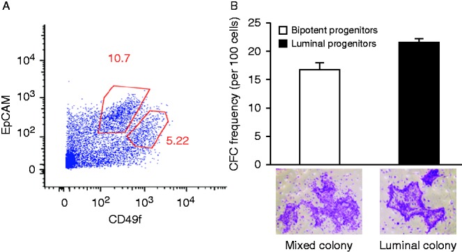
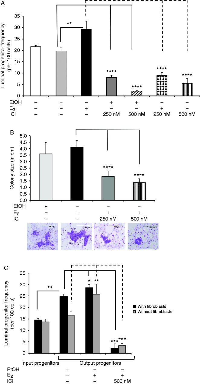
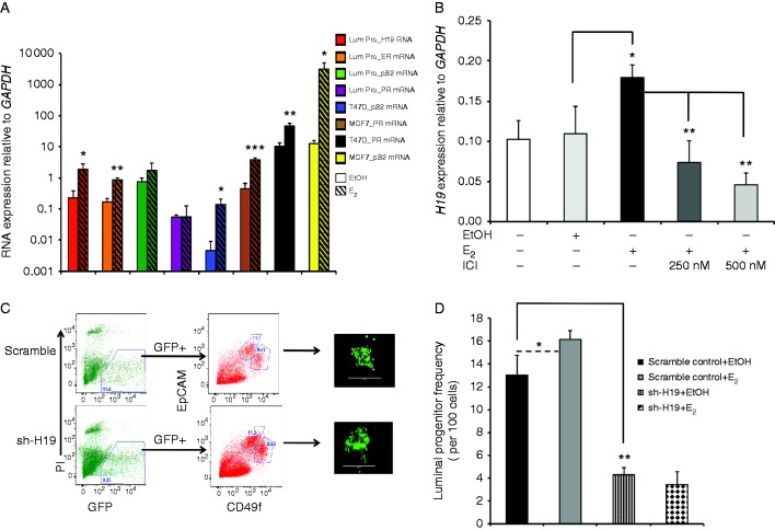
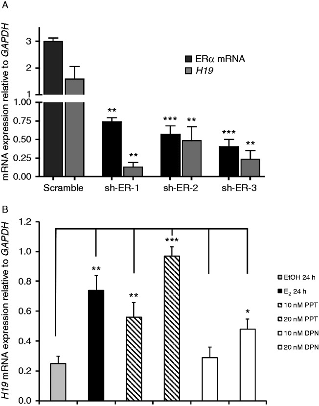
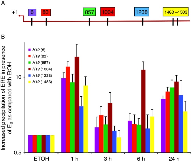
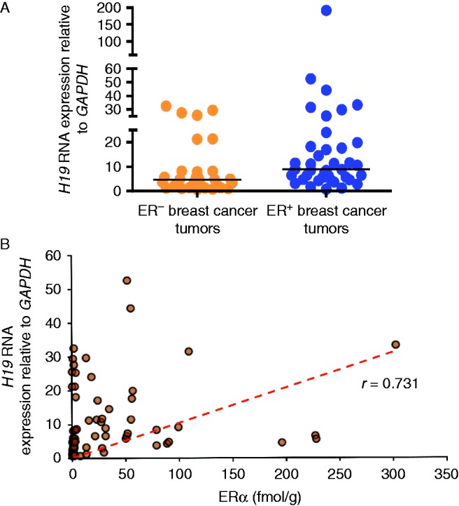
Although the role of estrogen signaling in breast cancer development has been extensively studied, the mechanisms that regulate the indispensable role of estrogen in normal mammary gland development have not been well studied. Because of the unavailability of culture system to maintain estrogen-receptor-positive (ERα(+)) cells in vitro, the molecular mechanisms that regulate estrogen/ERα signaling in the normal human breast are unknown. In the present study, we examined the effects of estrogen signaling on ERα(+) human luminal progenitors using a modified matrigel assay and found that estrogen signaling increased the expansion potential of these progenitors. Furthermore, we found that blocking ERα attenuated luminal progenitor expansion and decreased the luminal colony-forming potential of these progenitors. Additionally, blocking ERα decreased H19 expression in the luminal progenitors and led to the development of smaller luminal colonies. We further showed that knocking down the H19 gene in the luminal progenitors significantly decreased the colony-forming potential of the luminal progenitors, and this phenotype could not be rescued by the addition of estrogen. Lastly, we explored the clinical relevance of the estrogen-H19 signaling axis in breast tumors and found that ERα(+) tumors exhibited a higher expression of H19 as compared with ERα(-) tumors and that H19 expression showed a positive correlation with ERα expression in those tumors. Taken together, the present results indicate that the estrogen-ERα-H19 signaling axis plays a role in regulating the proliferation and differentiation potentials of the normal luminal progenitors and that this signaling network may also be important in the development of ER(+) breast cancer tumors.

Keywords: ER+ breast cancer cells; ERα; H19; luminal progenitors.


Figures

Figure 1
Figure 1
ERα is strongly expressed in luminal progenitors. (A) Luminal (EpCAMbrig ht CD49flow) and bipotent (EpCAMlowCD49fbright) progenitors were isolated from reduction mammoplasty samples using FACS. (B) CFC assays were used to determine the purity of different progenitor subtypes. As shown, 21.6% of the EpCAMbrightCD49flow cells formed pure luminal colonies, whereas 16.7% of the EpCAMlow CD49fbright cells formed mixed colonies (n=3). Representative luminal and mixed colonies are shown in the photographs.
Figure 2
Figure 2
Estrogen signaling enhances luminal progenitor proliferation. (A) Luminal progenitors were examined using CFC assays and were either treated with E2, EtOH, E2 plus ICI, or EtOH plus ICI at the indicated concentrations. The CFC cultures were fixed and stained, and the colony numbers were quantified (n=3). As shown, in the presence of E2, the colony-forming capacity of luminal progenitors was increased (1.48-fold as compared to EtOH), and blocking ERα significantly attenuated this induction. (B) Luminal progenitors were treated as in (A), and colony size (i.e., the ability of the luminal progenitors to form mature luminal cells) was determined by counting the number of cells in each colony. As shown, ICI treatment significantly reduced colony size in a dose-dependent manner. Images show representative colonies from each treatment. (C) Luminal progenitors were placed in matrigel cultures with or without irradiated fibroblasts (i3T3) in complete medium for 7 days and were then treated with E2, E2 plus ICI, or EtOH for an additional 7 days. Thereafter, the frequency of progenitors was assessed via CFC assay. In matrigels without i3T3, E2 significantly increased luminal progenitor frequency, which was circumvented with ICI. Interestingly, the addition of i3T3 alone increased progenitor cell numbers, and the addition of E2 had a small but statistically significant effect. Aliquots from the same samples were placed in the CFC assays before the matrigel cultures to give the starting luminal progenitor frequency. *P<0.05, **P<0.005, ***P<0.0001, ****P<0.00001.
Figure 3
Figure 3
Estrogen signaling enhances luminal progenitor proliferation through H19. (A) Luminal progenitors were placed in matrigel assays for 7 days and were then grown in estrogen-depleted growth media for 48 h and were subsequently treated with E2 (10 nM) or EtOH for an additional 24 h. qPCR was used to examine the expression of the estrogen target genes (PR, pS2, ERα, and H19) in cells obtained from the gels. Interestingly, no changes in the transcript levels of PR or pS2 could be detected. However, ERα and H19 expression levels were increased 5.3- and 8.5-fold respectively in E2-exposed luminal progenitors. For comparison, ER+ breast cancer cells (MCF7 and T47D) were exposed to EtOH or E2 for 24 h, and the expression of estrogen target genes was assessed using qPCR. Solid bars represent transcript expression in EtOH-treated cells, and crossed bars represent E2-treated cells. All of the transcript expression levels were normalized to the GAPDH levels, and the means and s.d.s are shown (n=3). (B) Luminal progenitors were isolated and placed in CFC assays; they were then treated with E2 (10 nM), E2 plus ICI, or EtOH for 7 days. H19 expression was examined via qPCR. As shown, in E2-stimulated colonies, H19 expression was increased 1.6-fold, whereas the addition of ICI significantly decreased H19 expression. (C) Lenti sh-H19 or lenti sh-scrambled-infected luminal progenitors (GFP+) were isolated via FACS and placed in CFC assays. Scale bars represent 400 μm. Representative pictures show the GFP+ colonies that developed from the transduced luminal progenitors. (D) Average colony counts were used as a prospective measure of the transduced luminal progenitor frequency (n=3). Compared with the scrambled control, the sh-H19-infected progenitors formed threefold fewer colonies, and the addition of E2 had no effect on these transduced progenitors. *P<0.05, **P<0.005, ***P<0.0005.
Figure 4
Figure 4
Estrogen-regulated expression of H19 is mainly regulated through ERα. (A) ERα transcript levels were decreased in MCF7 cells with three different sh-RNA fragments (sh-ER-1, -2, and -3) using lentiviral transduction. ERα and H19 transcript levels were examined in the transduced cells by qPCR. All of the data are normalized to the GAPDH transcript levels. (B) MCF7 cells were grown under estrogen-deprived growth conditions and treated with either EtOH, E2, PPT (a selective ERα agonist), or DPN (a selective ERβ agonist) for 24 h. H19 expression was ascertained using qPCR and was normalized with respect to GAPDH expression (n=3). Compared with EtOH, as little as 10 nm PPT increased H19 transcript levels, whereas DPN at the 10 nM concentration level had no effect. However, at 20 nM concentration, both PPT and DPN increased H19 expression (3.9- and 1.9-fold respectively). *P<0.05, **P<0.005, ***P<0.0005.
Figure 5
Figure 5
ERα binds to H19 promoter through EREs. (A) This figure shows the location of EREs within 1500 base pairs of the transcription start site (TSS, +1) of the H19 proximal promoter. Each box is representative of one ERE half-site, and the number within each box shows its location away from the TSS. (B) Chromatin immunoprecipitation was performed to examine the ERα occupancy of each ERE. MCF7 cells were grown in estrogen-depleted media and treated either with EtOH or E2 for the indicated times. ERα bound to each ERE was quantified using qPCR. As shown, ERα binding to the H19 promoter was detectable after 1 h of exposure to E2.
Figure 6
Figure 6
H19 expression is associated with ER+ breast tumors. (A) Expression of the H19 gene was quantified in 39 ER+ and 31 ER primary breast tumors using qPCR. On average, the ER+ breast tumors showed higher expression of H19 (mean±s.d.=17.65±31.04) as compared with the ER tumors (8.11±9.4) as per the Mann–Whitney test (P=0.0030). (B) Based on a ligand binding assay, the expression of ERα was found to range from 0 to 302 fmol/mg protein. Pearson correlation revealed a positive correlation between ERα and H19 expression in the breast tumors (r=0.731).

Similar articles

Cited by

References

    1. Adriaenssens E, Dumont L, Lottin S, Bolle D, Lepretre A, Delobelle A, Bouali F, Dugimont T, Coll J, Curgy JJ. H19 overexpression in breast adenocarcinoma stromal cells is associated with tumor values and steroid receptor status but independent of p53 and Ki-67 expression. American Journal of Pathology. 1998;153:1597–1607. doi: 10.1016/S0002-9440(10)65748-3. - DOI - PMC - PubMed
    1. Adriaenssens E, Lottin S, Dugimont T, Fauquette W, Coll J, Dupouy JP, Boilly B, Curgy JJ. Steroid hormones modulate H19 gene expression in both mammary gland and uterus. Oncogene. 1999;18:4460–4473. doi: 10.1038/sj.onc.1202819. - DOI - PubMed
    1. Adriaenssens E, Lottin S, Berteaux N, Hornez L, Fauquette W, Fafeur V, Peyrat JP, Le Bourhis X, Hondermarck H, Coll J, et al. Cross-talk between mesenchyme and epithelium increases H19 gene expression during scattering and morphogenesis of epithelial cells. Experimental Cell Research. 2002;275:215–229. doi: 10.1006/excr.2002.5500. - DOI - PubMed
    1. Anderson E, Clarke RB, Howell A. Estrogen responsiveness and control of normal human breast proliferation. Journal of Mammary Gland Biology and Neoplasia. 1998;3:23–35. doi: 10.1023/A:1018718117113. - DOI - PubMed
    1. Ariel I, Ayesh S, Perlman EJ, Pizov G, Tanos V, Schneider T, Erdmann VA, Podeh D, Komitowski D, Quasem AS, et al. The product of the imprinted H19 gene is an oncofetal RNA. Molecular Pathology. 1997;50:34–44. doi: 10.1136/mp.50.1.34. - DOI - PMC - PubMed

Publication types