. 2015 Jun 19;290(25):15475-15486.
doi: 10.1074/jbc.M115.636670. Epub 2015 May 5.

UDP-galactose (SLC35A2) and UDP-N-acetylglucosamine (SLC35A3) Transporters Form Glycosylation-related Complexes with Mannoside Acetylglucosaminyltransferases (Mgats)

Affiliations

UDP-galactose (SLC35A2) and UDP-N-acetylglucosamine (SLC35A3) Transporters Form Glycosylation-related Complexes with Mannoside Acetylglucosaminyltransferases (Mgats)

Dorota Maszczak-Seneczko et al. J Biol Chem. .

Abstract

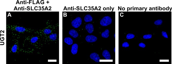
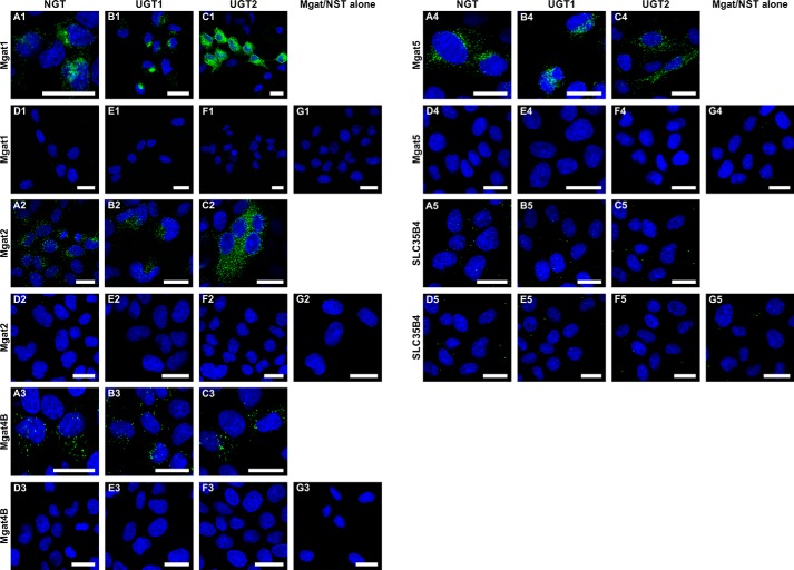
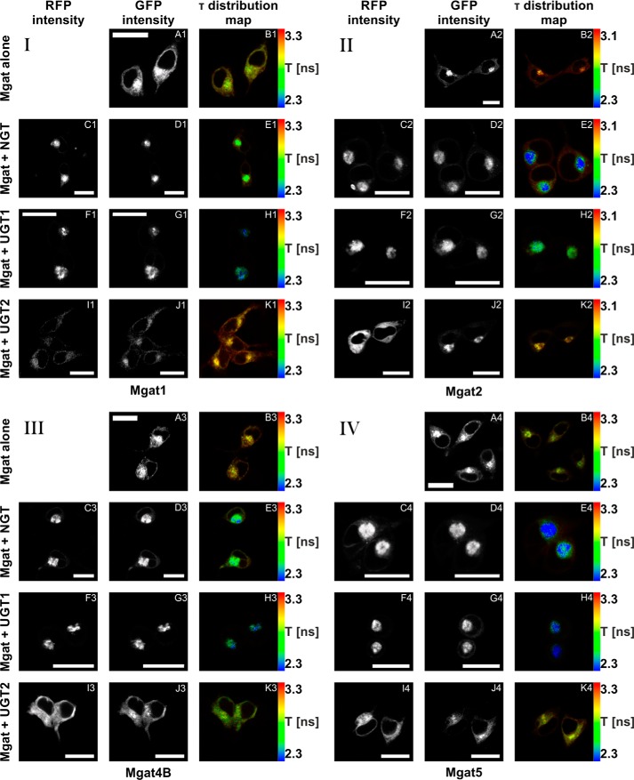
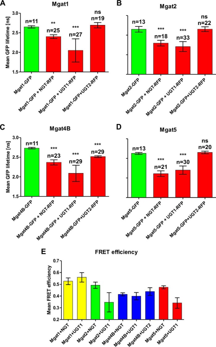
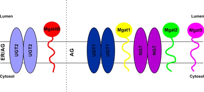
UDP-galactose transporter (UGT; SLC35A2) and UDP-N-acetylglucosamine transporter (NGT; SLC35A3) form heterologous complexes in the Golgi membrane. NGT occurs in close proximity to mannosyl (α-1,6-)-glycoprotein β-1,6-N-acetylglucosaminyltransferase (Mgat5). In this study we analyzed whether NGT and both splice variants of UGT (UGT1 and UGT2) are able to interact with four different mannoside acetylglucosaminyltransferases (Mgat1, Mgat2, Mgat4B, and Mgat5). Using an in situ proximity ligation assay, we found that all examined glycosyltransferases are in the vicinity of these UDP-sugar transporters both at the endogenous level and upon overexpression. This observation was confirmed via the FLIM-FRET approach for both NGT and UGT1 complexes with Mgats. This study reports for the first time close proximity between endogenous nucleotide sugar transporters and glycosyltransferases. We also observed that among all analyzed Mgats, only Mgat4B occurs in close proximity to UGT2, whereas the other three Mgats are more distant from UGT2, and it was only possible to visualize their vicinity using proximity ligation assay. This strongly suggests that the distance between these protein pairs is longer than 10 nm but at the same time shorter than 40 nm. This study adds to the understanding of glycosylation, one of the most important post-translational modifications, which affects the majority of macromolecules. Our research shows that complex formation between nucleotide sugar transporters and glycosyltransferases might be a more common phenomenon than previously thought.

Keywords: Golgi; UDP-N-acetylglucosamine transporter; UDP-galactose transporter; fluorescence resonance energy transfer (FRET); glycosylation; glycosyltransferase; mannoside acetylglucosaminyltransferase; protein complex; proximity ligation assay; sugar transport.


Figures

FIGURE 1.
FIGURE 1.
In situ PLA controls. A, PLA analysis of UGT2 N and C terminus proximity performed in MDCK-RCAr cells. B, negative control where anti-FLAG antibody was omitted during the PLA procedure. C, negative control where both primary antibodies were omitted during the PLA procedure. Bars, 20 μm.
FIGURE 2.
FIGURE 2.
Visualization of interactions between endogenous Mgats and UDP-sugar transporters using in situ PLA. A and B, PLA analysis of either Mgat1 (panels 1), Mgat2 (panels 2), Mgat4B (panels 3), or Mgat5 (panels 4) interaction with UGT (A) and NGT (B) in PC-3 cell line. C, negative control for Mgats-UDP-sugar transporters interaction analysis, where primary antibody against Mgat1/Mgat2/Mgat4B/Mgat5 was not added during the PLA procedure. Negative control for Mgat-UDP-sugar transporters interaction analysis, where primary antibody against UGT/NGT was not added during the PLA procedure (panels 5). D, negative control for Mgat-UDP-sugar transporters interaction where both primary antibodies were omitted during the PLA procedure. Bars, 20 μm.
FIGURE 3.
FIGURE 3.
Visualization of interactions between overexpressed Mgats and UDP-sugar transporters using in situ PLA. A–C, PLA analysis of either Mgat1 (panels 1), Mgat2 (panels 2), Mgat4B (panels 3), Mgat5 (panels 4), or SLC35B4 (panels 5) putative interaction with NGT (A), UGT1 (B), and UGT2 (C). D, negative control for HA-NGT and FLAG-Mgats/SLC35B4 interaction analysis, where primary antibodies were not added during the PLA procedure. E, negative control for HA-UGT1 and FLAG-Mgats/SLC35B4 interaction analysis, where primary antibodies were not added during the PLA procedure. F, negative control for HA-UGT2 and FLAG-Mgats/SLC35B4 interaction analysis, where primary antibodies were not added during the PLA procedure. G, negative control for HA-tagged UDP-sugar transporters and FLAG-Mgats/SLC35B4 interaction analysis, where MDCK-RCAr cells expressing only FLAG-Mgats/SLC35B4 were used, and anti-FLAG antibody was omitted during the PLA procedure. Bars, 20 μm.
FIGURE 4.
FIGURE 4.
In vivo FLIM-FRET analysis of interactions between Mgats and UDP-sugar transporters. A–K, confocal intensity-resolved (A, C, D, F, G, I, and J) and time-resolved (B, E, H, and K) imaging of eGFP-Mgat (D, G, and J) interaction with mRFP-UDP-sugar transporter (C, F, and I) in HEK293T cells in comparison with cells expressing eGFP-Mgat only (A). FLIM-FRET analysis of either Mgat1 (panel I), Mgat2 (panel II), Mgat4B (panel III), or Mgat5 (panel IV) putative interactions with NGT (C–E), UGT1 (F–H), and UGT2 (I–K). GFP fluorescence lifetime (τ) was shortened by simultaneous overexpression of eGFP-Mgats with both mRFP-NGT and mRFP-UGT1, strongly suggesting an interaction between these proteins. In the case of mRFP-UGT2, the same phenomenon was demonstrated only upon eGFP-Mgat4B co-expression. Co-expression of mRFP-UGT2 with eGFP-Mgat1, Mgat2, and Mgat5 did not influence GFP fluorescence lifetime. The red to blue color shift reflects shortening of the fluorescence lifetime. The rainbow scale bars placed next to time-resolved images (B, E, H, and K) represent fluorescence lifetime range between either 2.3 (blue) and 3.3 ns (red; Mgat1, Mgat4B, or Mgat5) or 2.3 (blue) and 3.1 ns (red; Mgat2). Bars, 20 μm. τ, fluorescence lifetime.
FIGURE 5.
FIGURE 5.
In vivo FLIM-FRET analysis of interactions between Mgats and UDP-sugar transporters. A–D, mean GFP lifetime values in the absence and in the presence of acceptor are shown. The data are shown as means ± S.D. from several measurements of the indicated cell number (n). Statistically significant (one-way analysis of variance test, p < 0.001) reduction of GFP lifetime upon co-expression of fluorophore-tagged Mgats and NGT, as well as UGT1, was demonstrated by comparing with eGFP-Mgats alone. No difference in GFP lifetime was observed when mRFP-UGT2 and eGFP-Mgat1, eGFP-Mgat2, and eGFP-Mgat5 were co-expressed. Only upon simultaneous expression of mRFP-UGT2 and eGFP-Mgat4B was a statistically significant decrease in GFP lifetime observed. E, mean FRET efficiency calculated for the FRET-positive Mgat-UDP-sugar transporter combinations. The data are shown as means ± S.D. from several measurements of the indicated (A–D) cell number (n). Mean FRET efficiency was calculated to estimate relative distances between interacting proteins (the higher the mean FRET efficiency, the shorter the distance between two proteins).
FIGURE 6.
FIGURE 6.
Schematic representation of complex formation between UDP-sugar transporters and Mgats. We hypothesize that in the ER UDP-sugar transporters recruit glycosyltransferase homodimers of one type at a time and accompany them on their way to the target Golgi compartment, where subsequent association of incoming NSTs brings distinct glycosyltransferase homodimers together, thus enabling a rearrangement that results in glycosyltransferase heterooligomerization. ER, endoplasmic reticulum; AG, Golgi apparatus.

References

    1. Hadley B., Maggioni A., Ashikov A., Day C. J., Haselhorst T., Tiralongo J. (2014) Structure and function of nucleotide sugar transporters: current progress. Comput. Struct. Biotechnol. J. 10, 23–32 - PMC - PubMed
    1. Coates S. W., Gurney T., Jr., Sommers L. W., Yeh M., Hirschberg C. B. (1980) Subcellular localization of sugar nucleotide synthetases. J. Biol. Chem. 255, 9225–9229 - PubMed
    1. Münster A. K., Eckhardt M., Potvin B., Mühlenhoff M., Stanley P., Gerardy-Schahn R. (1998) Mammalian cytidine 5′-monophosphate N-acetylneuraminic acid synthetase: a nuclear protein with evolutionarily conserved structural motifs. Proc. Natl. Acad. Sci. U.S.A. 95, 9140–9145 - PMC - PubMed
    1. Gerardy-Schahn R., Oelmann S., Bakker H. (2001) Nucleotide sugar transporters: Biological and functional aspects. Biochimie 83, 775–782 - PubMed
    1. Caffaro C. E., Hirschberg C. B. (2006) Nucleotide sugar transporters of the Golgi apparatus: from basic science to diseases. Acc. Chem. Res. 39, 805–812 - PubMed

Publication types

MeSH terms

Substances

LinkOut - more resources