. 2015 Jun 26;290(26):16202-14.
doi: 10.1074/jbc.M115.645978. Epub 2015 May 5.

FBXO32 Targets c-Myc for Proteasomal Degradation and Inhibits c-Myc Activity

Affiliations

FBXO32 Targets c-Myc for Proteasomal Degradation and Inhibits c-Myc Activity

Zhichao Mei et al. J Biol Chem. .

Abstract

FBXO32 (MAFbx/Atrogin-1) is an E3 ubiquitin ligase that is markedly up-regulated in muscle atrophy. Although some data indicate that FBXO32 may play an important role in tumorigenesis, the molecular mechanism of FBXO32 in tumorigenesis has been poorly understood. Here, we present evidence that FBXO32 targets the oncogenic protein c-Myc for ubiquitination and degradation through the proteasome pathway. Phosphorylation of c-Myc at Thr-58 and Ser-62 is dispensable for FBXO32 to induce c-Myc degradation. Mutation of the lysine 326 in c-Myc reduces c-Myc ubiquitination and prevents the c-Myc degradation induced by FBXO32. Furthermore, overexpression of FBXO32 suppresses c-Myc activity and inhibits cell growth, but knockdown of FBXO32 enhances c-Myc activity and promotes cell growth. Finally, we show that FBXO32 is a direct downstream target of c-Myc, highlighting a negative feedback regulation loop between c-Myc and FBXO32. Thus, FBXO32 may function by targeting c-Myc. This work explains the function of FBXO32 and highlights its mechanisms in tumorigenesis.

Keywords: FBXO32; Myc (c-Myc); cell proliferation; muscle atrophy; oncogene; proteasomal degradation; ubiquitylation (ubiquitination).


Figures

FIGURE 1.
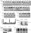
FIGURE 1.
FBXO32 promotes c-Myc turnover. A, ectopic expression of HA-FBXO32 induced degradation of ectopic expressed HA-c-Myc. HEK293T cells were transfected with equal amounts of HA-c-Myc along with increasing amounts of HA-FBXO32, compensated with a CMV-HA empty vector to keep the same amount of transfected plasmid DNA. Quantization of the protein levels is shown on the right. B, knockdown of FBXO32 by shRNAs causes the protein levels of endogenous c-Myc and its target E2F2 to be increased in HCT116 cells. Quantification of the protein levels is shown on the right. C, ectopic expression of FBXO32-shRNA-2-resistant FBXO32 cDNA can rescue the effect of FBXO32-shRNA-2 on the endogenous protein level of c-Myc in HCT116 cells. Quantization of the protein levels is shown on the right. D, co-transfection of HA-FBXO32 enhances c-Myc protein instability in the presence of protein synthesis inhibitor CHX (50 μg/ml), but co-transfection of HA-FBXO32-ΔF-box (F-box deleted) does not. HCT116 cells were transiently transfected with the indicated plasmids. After 24 h, CHX was added to the cells, and cell lysates were prepared at the indicated time points. Quantization of the protein levels is shown on the right. E, knockdown of FBXO32 by FBXO32-shRNA-1 or FBXO32-shRNA-1 in HCT116 cells enhances endogenous c-Myc protein stability in the presence of protein synthesis inhibitor CHX (50 μg/ml). Quantization of the protein levels is shown on the right. Multi Gauge version 3.0 was used for quantifying protein levels based on band density obtained in Western blot assays; the statistical analysis was performed using GraphPad Prism version 5.0 (unpaired Student's t tests). Error bars, S.E.
FIGURE 2.
FIGURE 2.
FBXO32-mediated c-Myc degradation is independent of the phosphorylation of c-Myc at Thr-58 and Ser-62. A, co-transfection of HA-FBXO32 causes protein degradation of wild-type c-Myc as well as c-Myc mutants T58A, S62S, and T58A/S62A. B, co-transfection of HA-FBXO32 causes protein degradation of wild-type c-Myc as well as c-Myc mutants P57S and S62E. C, co-transfection of HA-FBXO32 causes protein degradation of wild-type c-Myc as well as c-Myc mutants S71A, Y74A, T358A, and S373A.
FIGURE 3.
FIGURE 3.
FBXO32 directly interacts with c-Myc. A, co-immunoprecipitation assays show FLAG-tagged FBXO32 interacts with HA-tagged c-Myc when they are overexpressed in HEK293T cells. B, endogenous FBXO32 interacts with endogenous c-Myc, as revealed by co-immunoprecipitation assays using anti-c-Myc antibody in mouse muscle lysates. Rabbit IgG is used as control for co-immunoprecipitation assays. C, bacterial expressed GST-tagged FBXO32 directly interacts with bacterial expressed His-tagged c-Myc. Bacterial expressed GST protein is used as control. D, the wild-type c-Myc as well as its mutants T58A, S62A, and T58A/S62A interact with FBXO32, as revealed by co-immunoprecipitation assays. E, schematic of the c-Myc domains. The extent of the interaction between FBXO32 and the c-Myc domains is indicated by the number of plus signs. F and G, co-immunoprecipitation of human FLAG-FBXO32 with HA-tagged c-Myc domains in HEK293T cells transfected with the indicated plasmids. H, schematic of the FBXO32 domains. The extent of the interaction between c-Myc and the FBXO32 domains is indicated by the number of plus signs. I, co-immunoprecipitation of human HA-c-Myc with FLAG-tagged FBXO32 domains in HEK293T cells transfected with the indicated plasmids. J, co-immunoprecipitation of human HA-c-Myc with GFP-tagged FBXO32 domains in HEK293T cells transfected with the indicated plasmids. IP, immunoprecipitation; IB, immunoblot; n.s., nonspecific; TCL, total cell lysate.
FIGURE 4.
FIGURE 4.
FBXO32 catalyzes c-Myc for Lys-48-linked ubiquitination and proteasomal degradation. A, the proteasome inhibitor MG132 blocks FBXO32-induced c-Myc degradation. HEK293T cells were transfected with HA-c-Myc together with either HA-FBXO32 or empty vector; MG132 (20 μm) was added to the medium 6 h before protein harvest. B, the wild-type FBXO32 (FLAG-FBXO32-WT) catalyzes c-Myc ubiquitination, but when the F-box is deleted, the mutant (FLAG-FBXO32-ΔF-Box) reduces its catalytic capability dramatically for c-Myc ubiquitination. HEK293T cells were transfected with HA-c-Myc and His-Ub-WT or alone with FLAG-FBXO32-WT or FLAG-FBXO32-ΔF-box; 24 h after transfection, lysates were prepared and subjected to immunoprecipitation by Ni2+-nitrilotriacetic acid beads and then were detected by Western blot using anti-HA antibody. C, the wild-type FBXO32 (FLAG-FBXO32-WT) catalyzes c-Myc for Lys-48-linked ubiquitination but not for Lys-63-linked ubiquitination. HEK293T cells were transfected with HA-c-Myc and FLAG-FBXO32 or alone with His-tagged wild-type ubiquitin (His-Ub-WT), His-tagged ubiquitin 48 Lys/Arg mutant (Ub-K48R), or His-tagged ubiquitin 63 Lys/Arg mutant (His-Ub-K63R); the ubiquitination assays were performed as in B. D, Lys-48-linked ubiquitination catalyzed by the wild-type FBXO32 (FLAG-FBXO32-WT) was further confirmed by seven Lys-only ubiquitin mutants, Lys-6, -11, -27, -29, -33, -48, and -63. IP, immunoprecipitation; IB, immunoblot.
FIGURE 5.
FIGURE 5.
FBXO32 targets c-Myc ubiquitination at K326. A, HEK293T cells were transfected with the indicated c-Myc mutants together with either FBXO32 or empty vector. The expressions of c-Myc were detected by Western blot analysis using anti-HA antibody. B, degradation of the c-Myc mutants, K51R, K52R, and K51R/K52R induced by FBXO32 was further confirmed. C, degradation of the c-Myc multiple mutant, K8R, was further confirmed. D, protein levels were quantified based on band density obtained in Western blot assays; the protein level with HA empty vector transfection was treated as 1; the statistical analysis was performed using GraphPad Prism version 5.0 (unpaired Student's t tests); the protein level with HA empty vector transfection was treated as 1. E, the catalytic capability of FBXO32 on c-Myc(Lys-326) polyubiquitination is reduced significantly (the sixth column from the left to the right) compared with that on wild-type c-Myc (the second column from the left to the right). F, the c-Myc Lys/Arg mutant (HA-c-Myc(K326R)), as well as the wild-type c-Myc (HA-c-Myc(WT)), interacts with FBXO32, as revealed by co-immunoprecipitation assays. For ectopic expression, the amount of HA-c-Myc(WT) was 2 times more than that of HA-c-Myc(K326R) (ratio = 1:3) regarding the degradation of wild-type c-Myc by FBXO32. G, the catalytic capability of FBXO32 on the c-Myc multiple mutant (K326R plus K8R) polyubiquitination is further reduced (the eighth column from the left to the right) compared with that on the c-Myc(K326R) (the fourth column from the left to the right). Error bars, S.E. IP, immunoprecipitation; IB, immunoblot.
FIGURE 6.
FIGURE 6.
FBXO32 inhibits c-Myc transcriptional activity. A, overexpression of FBXO32 enhances the activity of Gadd45α promoter reporter activity suppressed by c-Myc (p = 0.0044) in HEK293T cells. B and C, overexpression of FBXO32 suppresses the activity of E2F2 wild-type promoter reporter activity induced by c-Myc (p = 0.0190) but not the activity of E2F2 promoter mutated reporter in HEK293T cells. D, the expressions of transfected HA-FBXO32 and HA-c-Myc in HEK293T cells are confirmed by Western blot using anti-HA antibody. E, the mRNA level of E2F2 is reduced dramatically when FBXO32 is overexpressed by the lentivirus infection expressing FBXO32 (Phage-FBXO32) compared with the control with the lentivirus infection expressing GFP protein in A673 cells. F, the mRNA level of Gadd45α is increased dramatically when FBXO32 is overexpressed by the lentivirus infection expressing FBXO32 (Phage-FBXO32) compared with the control with the lentivirus infection expressing GFP protein in A673 cells. G, the mRNA level of E2F2 is increased dramatically when FBXO32 is knocked down by the lentivirus infection expressing FBXO32-shRNA-1 and FBXO32-shRNA-2 compared with the control with the lentivirus infection expressing GFP-shRNA in A673 cells. H, the mRNA level of Gadd45α is reduced dramatically when FBXO32 is knocked down by the lentivirus infection expressing FBXO32-shRNA-1 and FBXO32-shRNA-2 compared with the control with the lentivirus infection expressing GFP-shRNA in A673 cells. I, overexpression of FBXO32 by lentivirus infection has no effect on Gadd45α promoter reporter activity in c-Myc-knocked down HCT116 cells (p = 0.1051). J, when c-Myc(WT) is re-expressed in c-Myc-knocked down HCT116 cells by lentivirus infection, the enhancement effect of FBXO32 on the activity of Gadd45α promoter reporter activity suppressed by c-Myc is restored (p = 0.0075). K, when the mutated c-Myc(K326R) is re-expressed in c-Myc-knocked down HCT116 cells by lentivirus infection, the enhancement effect of FBXO32 on the activity of Gadd45α promoter reporter activity suppressed by c-Myc is still not restored (p = 0.1115). L, the expressions of the HA-tagged wild-type c-Myc, the HA-tagged mutated c-Myc(K326R), and the HA-tagged FBXO32 by lentivirus infection in HCT116 cells are confirmed by Western blot analysis. M, overexpression of FBXO32 by transient transfection has no effect on E2F2 promoter reporter activity in c-Myc-knocked down HCT116 cells (p = 0.4194). N, when c-Myc(WT) is re-expressed in c-Myc-knocked down HCT116 cells by lentivirus infection, the inhibitory effect of FBXO32 on the activity of Gadd45α promoter reporter activity up-regulated by c-Myc is restored (p = 0.0002). ○, when the mutated c-Myc(K326R) is re-expressed in c-Myc-knocked down HCT116 cells by lentivirus infection, the inhibitory effect of FBXO32 on the activity of Gadd45α promoter reporter activity up-regulated by c-Myc is still not restored (p = 0.1325). P, overexpression of FBXO32 by transient transfection has no effect on mutated E2F2 promoter reporter (E2F-mut-luc) activity in c-Myc-knocked down HCT116 cells (p = 0.1325). Q, when the wild-type c-Myc(WT) is re-expressed in c-Myc-knocked down HCT116 cells by lentivirus infection, the inhibitory effect of FBXO32 on the activity of mutated E2F2 promoter reporter activity up-regulated by c-Myc is still restored (p = 0.3658). Data are presented as mean ± S.E. (error bars) of three independent experiments performed in triplicate; the statistical analysis was performed using GraphPad Prism version 5.0 (unpaired Student's t tests). IB, immunoblot.
FIGURE 7.
FIGURE 7.
FBXO32 inhibits ovary cancer cell SKOV3 proliferation. A, overexpression of wild-type FBXO32 by lentivirus infection in SKOV3 cells inhibits cell proliferation significantly after day 2 compared with the control, but overexpression of F-box-deleted mutant of FBXO32 (FBXO32-ΔF-box) has no obvious effect. The SKOV3 cells transduced with lentivirus vectors stably expressing GFP and FBXO32 or GFP alone (as a control) were seeded in 6-well plates with 8 × 10³ cells/well. B, knockdown of FBXO32 by FBXO32-shRNA-1 or FBXO32-shRNA-2 in SKOV3 cells enhances cell proliferation after day 2 compared with the control. The SKOV3 cells transduced with lentivirus vectors stably expressing FBXO32-shRNA-1, FBXO32-shRNA-2, or GFP-shRNA (as a control) were seeded in 6-well plates with 8 × 10³ cells/well. The cell numbers were counted every 2 days using an automated cell counter (Bio-Rad, TC20TM). C, overexpression of wild-type FBXO32 inhibits colony formation, but overexpression of F-box-deleted mutant of FBXO32 (FBXO32-ΔF-box) has no obvious effect. Data are presented as mean ± S.E. (error bars) of three independent experiments performed in triplicate; the statistical analysis was performed using GraphPad Prism version 5.0 (t tests).
FIGURE 8.
FIGURE 8.
FBXO32 is a direct target of c-Myc. A, overexpression of c-Myc activates FBXO32 promoter reporter activity. A schematic of the FBXO32 promoter reporter constructs is shown on the left. B, the expression of HA-c-Myc is confirmed by Western blot analysis. C, schematic of one potential c-Myc binding site (E-box: CACGTG (in boldface type)) localized at the FBXO32 promoter (−204 to −5) and the mutated E-box construct. D, overexpression of c-Myc activates the wild-type FBXO32 promoter reporter (−204 to −5) activity but not the mutated FBXO32 promoter reporter activity. E, the mRNA level of FBXO32 is elevated when c-Myc is overexpressed in HEK293T cells, as revealed by a semiquantitative RT-PCR assay (p = 0.0061). F, the mRNA level of FBXO32 is elevated when c-Myc is overexpressed in HCT116 cells, as revealed by semiquantitative RT-PCR assays (p = 0.0003). G, the mRNA level of FBXO32 is reduced when c-Myc is knocked down in HCT116 cells by c-Myc-shRNA-1 and c-Myc-shRNA-2, as revealed by semiquantitative RT-PCR assays. H, the protein level of FBXO32 is elevated when c-Myc is overexpressed in HCT116 cells, as revealed by Western blot analysis using anti-c-Myc antibody. I, the protein level of FBXO32 is reduced when c-Myc is knocked down in HCT116 cells by c-Myc-shRNA-1 and c-Myc-shRNA-2 as revealed by Western blot analysis. J, schematic of locations of fragments amplified in FBXO32 promoter or β-actin promoter. K, ChIP analysis indicates that c-Myc interacts directly with the FBXO32 promoter region harboring the E-box. L, semiquantitative RT-PCR analysis confirms that c-Myc binds to the −204 to −5 region of the FBXO32 promoter. Data are presented as the means ± S.E. (error bars) of three independent experiments performed in triplicate.

Similar articles

Cited by

References

    1. Bodine S. C., Latres E., Baumhueter S., Lai V. K. M., Nunez L., Clarke B. A., Poueymirou W. T., Panaro F. J., Na E. Q., Dharmarajan K., Pan Z. Q., Valenzuela D. M., DeChiara T. M., Stitt T. N., Yancopoulos G. D., Glass D. J. (2001) Identification of ubiquitin ligases required for skeletal muscle atrophy. Science 294, 1704–1708 - PubMed
    1. Gomes M. D., Lecker S. H., Jagoe R. T., Navon A., Goldberg A. L. (2001) Atrogin-1, a muscle-specific F-box protein highly expressed during muscle atrophy. Proc. Natl. Acad. Sci. U.S.A. 98, 14440–14445 - PMC - PubMed
    1. Ponting C. P., Phillips C., Davies K. E., Blake D. J. (1997) PDZ domains: targeting signalling molecules to sub-membranous sites. Bioessays 19, 469–479 - PubMed
    1. Harrison S. C. (1996) Peptide-surface association: the case of PDZ and PTB domains. Cell 86, 341–343 - PubMed
    1. Julie L. C., Sabrina B. P., Marie-Pierre L., Leibovitch S. A. (2012) Identification of essential sequences for cellular localization in the muscle-specific ubiquitin E3 ligase MAFbx/Atrogin 1. FEBS Lett. 586, 362–367 - PubMed

Publication types

MeSH terms

Substances

LinkOut - more resources