. 2015:2015:247357.
doi: 10.1155/2015/247357. Epub 2015 Apr 6.

Aqueous Date Flesh or Pits Extract Attenuates Liver Fibrosis via Suppression of Hepatic Stellate Cell Activation and Reduction of Inflammatory Cytokines, Transforming Growth Factor- β 1 and Angiogenic Markers in Carbon Tetrachloride-Intoxicated Rats

Affiliations

Aqueous Date Flesh or Pits Extract Attenuates Liver Fibrosis via Suppression of Hepatic Stellate Cell Activation and Reduction of Inflammatory Cytokines, Transforming Growth Factor- β 1 and Angiogenic Markers in Carbon Tetrachloride-Intoxicated Rats

Nouf M Al-Rasheed et al. Evid Based Complement Alternat Med. 2015.

Abstract

Previous data indicated the protective effect of date fruit extract on oxidative damage in rat liver. However, the hepatoprotective effects via other mechanisms have not been investigated. This study was performed to evaluate the antifibrotic effect of date flesh extract (DFE) or date pits extract (DPE) via inactivation of hepatic stellate cells (HSCs), reducing the levels of inflammatory, fibrotic and angiogenic markers. Coffee was used as reference hepatoprotective agent. Liver fibrosis was induced by injection of CCl4 (0.4 mL/kg) three times weekly for 8 weeks. DFE, DPE (6 mL/kg), coffee (300 mg/kg), and combination of coffee + DFE and coffee + DPE were given to CCl4-intoxicated rats daily for 8 weeks. DFE, DPE, and their combination with coffee attenuated the elevated levels of inflammatory cytokines including tumor necrosis factor-α, interleukin-6, and interleukin-1β. The increased levels of transforming growth factor-β1 and collagen deposition in injured liver were alleviated by both extracts. CCl4-induced expression of α-smooth muscle actin was suppressed indicating HSCs inactivation. Increased angiogenesis was ameliorated as revealed by reduced levels and expression of vascular endothelial growth factor and CD31. We concluded that DFE or DPE could protect liver via different mechanisms. The combination of coffee with DFE or DPE may enhance its antifibrotic effects.


Figures

Figure 1
Figure 1
Light photomicrograph of liver sections stained with Masson's trichrome to demonstrate the fibrous tissue (scale bar 50 μm). (a) Control liver showing normal amount and distribution of fibrous tissue (arrow) mainly in the portal area. (b) Liver section from rat exposed to CCl4 showing increase of the fibrous tissue (arrow) which extends outside the portal area. (c) Liver from rat exposed to CCl4 and receiving 2 mL/kg of DFE showing mild decrease of the amount abnormal deposited fibrous tissue. Also (d) represents liver from rat exposed to CCl4 and 4 mL/kg of DFE and shows decrease of fibrous tissue but still abnormally deposited outside portal area. ((e), (f)) Liver sections from rat exposed to CCl4 and receiving 6 and 8 mL/kg of DFE, respectively, show apparently normal amount and distribution of fibrous tissue if compared with the control group (a).
Figure 2
Figure 2
Light photomicrographs of liver sections stained with Masson are trichrome to demonstrate the fibrous tissue (scale bar 50 μm). (a) Control liver showing normal amount and distribution of fibrous tissue (arrow) mainly in the portal area. (b) Liver section from rat exposed to CCl4 showing increase of the fibrous tissue (arrow) which extends outside the portal area. (c) Liver from rat exposed to CCl4 and receiving 2 mL/kg of DPE showing mild decrease of the amount abnormal deposited fibrous tissue. Also (d) represents liver from rat exposed to CCl4 and 4 mL/kg of DPE and shows decrease of fibrous tissue but still abnormally deposited outside portal area. ((e), (f)) Liver sections from rat exposed to CCl4 and receiving doses of 6 and 8 mL/kg of DPE, respectively, show apparently normal amount and distribution of fibrous tissue if compared with the control group (a).
Figure 3
Figure 3
Effect of coffee, date flesh extract (DFE), date pits extract (DPE), and the combination groups on hepatic levels of MDA, a marker of lipid peroxidation in CCl4-intoxicated rats. Values are expressed as mean ± SEM. a: significantly different from normal control group, b: significantly different from CCl4-treated group; c: significantly different from coffee-treated group, ∗∗∗ P < 0.001, ∗∗ P < 0.01, P < 0.05.
Figure 4
Figure 4
Effect of coffee, date flesh extract (DFE), date pits extract (DPE), and the combination groups on hepatic levels of reduced glutathione (GSH), glutathione reductase (GR), glutathione peroxidase (GPx) and superoxide dismutase (SOD) activities in CCl4-intoxicated rats. Values are expressed as mean ± SEM. a: significantly different from normal control group; b: significantly different from CCl4-treated group; c: significantly different from coffee-treated group; e: significantly different from DPE-treated group. ∗∗∗ P < 0.001, ∗∗ P < 0.01, P < 0.05.
Figure 5
Figure 5
Effect of date flesh extract (DFE), date pits extract (DPE), coffee, and the combination groups on hepatic levels of proinflammatory mediators. (a) Tumor necrosis factor α (TNF-α), (b) interleukin-6 (IL-6), and (c) interleukin-1β (IL-1β) in CCl4-intoxicated rats. Values are expressed as mean ± SEM. a: significantly different from normal control group; b: significantly different from CCl4-treated group; c: significantly different from coffee-treated group; d: significantly different from DFE-treated group. ∗∗∗ P < 0.001, ∗∗ P < 0.01, P < 0.05.
Figure 6
Figure 6
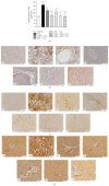
(a) Effect of date flesh extract (DFE), date pits extract (DPE), coffee and the combination groups on hydroxyproline content in hepatic tissue of CCl4-intoxicated rats. Values are expressed as mean ± SEM. a: significantly different from normal control group; b: significantly different from CCl4-treated group. ∗∗∗ P < 0.001, ∗∗ P < 0.01, P < 0.05. (b) Light microscopic photomicrographs of liver tissue stained with Masson's trichrome stain (scale bar = 50 μm). (A) Control liver showing normal hepatic fibrous tissue distribution restricted mainly to the portal area, while demarcation between classic hepatic lobule is observed due to very delicate fibrous tissue. (B) Represent liver section of rat receiving CCl4 showing marked increase of fibrous tissue arranged in irregular way between degenerated cells and sinusoids, causing destruction of classic architecture of hepatic lobules. Liver sections from rats treated with coffee (C), DFE (D), DPE (E), and the combination of coffee + DFE (F) and coffee + DPE (G) showing marked decrease of abnormally extra deposited fibrous tissue. The improvement is prominent in DFE than coffee alone or DPE alone. Sections from rats receiving combination showed apparently normal amount and distribution of fibrous tissue. (c) Effect of date flesh extract (DFE), date pits extract (DPE), coffee, and the combination groups on hepatic levels of transforming growth factor beta-1 (TGF-β1) in CCl4-intoxicated rats. Values are expressed as mean ± SEM. a: significantly different from normal control group; b: significantly different from CCl4-treated group; c: significantly different from coffee-treated group. ∗∗∗ P < 0.001, ∗∗ P < 0.01, P < 0.05. (d) Light microscopic photomicrographs of liver tissue immunostained with primary anti-TGF-β1 antibody (scale bar = 50 μm). (A) Control liver showing normal very weak immune reactivity of hepatocytes cytoplasm while the nuclei are not stained, and also the endothelial cells of both central vein and hepatic sinusoids are not stained. (B) represents liver section of rat receiving CCl4 showing strong abnormal immune reactivity of most of hepatocytes' cytoplasm and nuclei, and also most of cells of structures of portal area show strong nuclear immune reactivity. Panels (C), (D), (E), (F), and (G) represent liver sections from rat receiving coffee, DFE, DPE, coffee + DFE, and coffee + DPE, respectively, showing few hepatocytes with strong ((C), (E)) or moderate (D) immune reactivity especially those around portal area. In (F) and (G) hepatocytes' cytoplasm and few cells of structures of portal areas show weak positivity especially in rats receiving a combination of coffee + DFE (F). (e) Light microscopic photomicrographs of liver tissue immunostained with primary anti-α-SMA antibody (scale bar = 50 μm). (A) Control liver showing normal positive immune reactivity of smooth muscle of the blood vessels of the portal area without immune staining positivity in between hepatocytes, while (B) represents liver section of rat receiving CCl4 in which strong abnormal distributed immune reactivity, especially around degenerated hepatocytes, is prominent. Panels (C), (D), (E), (F), and (G) represent liver sections from rat receiving coffee, DFE, DPE, coffee + DFE, and coffee + DPE, respectively, showing marked decrease of the immune reactivity outside portal areas especially in groups receiving DFE and the combination.
Figure 7
Figure 7
(a) Effect of date flesh extract (DFE), date pits extract (DPE), coffee, and the combination groups on hepatic levels of vascular endothelial growth factor (VEGF) in CCl4-intoxicated rats. Values are expressed as mean ± SEM. a: significantly different from normal control group; b: significantly different from CCl4-treated group; c: significantly different from coffee-treated group. ∗∗∗ P < 0.001, ∗∗ P < 0.01, P < 0.05. (b) Light microscopic photomicrographs of liver tissue immunostained with primary anti-VGEF antibody (scale bar = 50 μm). (A) Control liver showing negative immune reactivity of hepatocytes, while (B) represents liver section of rat receiving CCl4 showing strong abnormal irregularly distributed immune reactivity, especially between degenerated hepatocytes' cytoplasm and nuclei. Panels (C), (D), (E), (F), and (G) that represent liver sections from rat receiving coffee, DFE, DPE, coffee + DFE, and coffee + DPE, respectively, showed marked decrease in the intensity of the immune reactivity of hepatocytes surrounding portal areas especially in rats treated with the DFE and the combination of coffee + DFE. (c) Light microscopic photomicrographs of liver tissue immunostained with anti-VEGFR-1 primary antibody (scale bar = 100 μm). (A) Control liver showing normal few weak immune positive cellular and nuclear receptors. (B) represents liver section of rat receiving CCl4 in which there are focal strong abnormal immune reactivity of most of hepatocytes' cell membrane and nuclei. Panels (C), (D), (E), (F), and (G) that represent liver sections from rat receiving coffee, DFE, DPE, coffee + DFE, and coffee + DPE, respectively, revealed a decrease in the number of immunostained hepatocytes with marked depletion of immune positivity observed in rats treated with coffee + DPE and then with coffee + DFE and DFE alone-treated groups. (d) Light microscopic photomicrographs of liver tissue immunostained with primary anti-PECAM (CD31) antibody (scale bar = 50 μm). (A) Control liver showing normal positive immune reactivity of endothelial cells cell membranes of the blood vessels of the portal area and blood sinusoids, while (B) represents liver section of rat receiving CCl4 in which strong abnormal irregularly distributed immune reactivity, especially around degenerated hepatocytes, is prominent. Panels (C), (D), (E), (F), and (G) that represent liver sections from rat treated with coffee, DFE, DPE, coffee + DFE, and coffee + DPE, respectively, showed marked decrease of the immune reactivity outside portal areas especially in groups receiving DFE and combination.
Figure 8
Figure 8
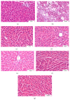
Light microscopic photomicrographs of liver tissue stained H&E (scale bar = 50 μm except B = 100 μm). (a) Control liver showing normal hepatic architecture, normal hepatocytes, and blood sinusoids, while (b) represents liver section of rat receiving CCl4 in which most of hepatocytes are degenerated and swollen with vacuolated cytoplasm and pyknotic nuclei. There is complete loss of hepatic lobule architecture. Panels (c), (d), (e), (f), and (g) represent liver sections from rats treated with coffee, DFE, DPE, and the combination of coffee + DFE and coffee + DPE, respectively, showing marked regeneration of hepatic architecture and almost all hepatocytes become normal especially rats receiving DFE alone or combinations.

Similar articles

Cited by

References

    1. Lee U. E., Friedman S. L. Mechanisms of hepatic fibrogenesis. Best Practice & Research Clinical Gastroenterology. 2011;25(2):195–206. doi: 10.1016/j.bpg.2011.02.005. - DOI - PMC - PubMed
    1. Ginès P., Cárdenas A., Arroyo V., Rodés J. Management of cirrhosis and ascites. The New England Journal of Medicine. 2004;350(16):1646–1654. doi: 10.1056/nejmra035021. - DOI - PubMed
    1. Kisseleva T., Brenner D. A. Hepatic stellate cells and the reversal of fibrosis. Journal of Gastroenterology and Hepatology. 2006;21(supplement 3):S84–S87. doi: 10.1111/j.1440-1746.2006.04584.x. - DOI - PubMed
    1. Friedman S. L. Mechanisms of hepatic fibrogenesis. Gastroenterology. 2008;134(6):1655–1669. doi: 10.1053/j.gastro.2008.03.003. - DOI - PMC - PubMed
    1. Gressner A. M., Weiskirchen R. Modern pathogenetic concepts of liver fibrosis suggest stellate cells and TGF-β as major players and therapeutic targets. Journal of Cellular and Molecular Medicine. 2006;10(1):76–99. doi: 10.1111/j.1582-4934.2006.tb00292.x. - DOI - PMC - PubMed

LinkOut - more resources