

The Roles of Growth Factors in Keratinocyte Migration
- PMID: 25945284
- PMCID: PMC4397993
- DOI: 10.1089/wound.2014.0540
The Roles of Growth Factors in Keratinocyte Migration
Abstract
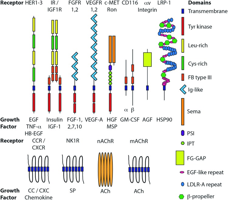
Significance: The re-epithelialization of wounded skin requires the rapid and coordinated migration of keratinocytes (KC) into the wound bed. Almost immediately after wounding, cells present at or attracted to the wound site begin to secrete a complex milieu of growth factors. These growth factors exert mitogenic and motogenic effects on KCs, inducing the rapid proliferation and migration of KCs at the wound edge. Recent Advances: New roles for growth factors in KC biology are currently being discovered and investigated. This review will highlight the growth factors, particularly transforming growth factor-α (TGF-α), heparin-binding epidermal growth factor (HB-EGF), insulin-like growth factor 1 (IGF-1), fibroblast growth factor 7 (FGF-7), FGF-10, and hepatocyte growth factor (HGF), which have conclusively been shown to be the most motogenic for KCs. Critical Issues: The cellular and molecular heterogeneity of wounded tissue makes establishing direct relationships between specific growth factors and KC migration difficult in situ. The absence of this complexity in simplified in vitro experimental models of migration makes the clinical relevance of the results obtained from these in vitro studies ambiguous. Future Directions: Deciphering the relationship between growth factors and KC migration is critical for understanding the process of wound healing in normal and disease states. Insights into the basic science of the effects of growth factors on KC migration will hopefully lead to the development of new therapies to treat acute and chronic wounds.
Figures


References
-
- Brown GL, Nanney LB, Griffen J, et al. . Enhancement of wound healing by topical treatment with epidermal growth factor. N Engl J Med 1989;321:76–79 - PubMed
-
- Uchi H, Igarashi A, Urabe K, et al. . Clinical efficacy of basic fibroblast growth factor (bFGF) for diabetic ulcer. Eur J Dermatol EJD 2009;19:461–468 - PubMed
-
- Fan K, Tang J, Escandon J, Kirsner RS. State of the art in topical wound-healing products. Plast Reconstruct Surg 2011;127 Suppl 1:44S–59S - PubMed
-
- Phillips T, Stanton B, Provan A, Lew R. A study of the impact of leg ulcers on quality of life: financial, social, and psychologic implications. J Am Acad Dermatol 1994;31:49–53 - PubMed
Publication types
LinkOut - more resources
Full Text Sources
Other Literature Sources
Miscellaneous
