. 2015 Apr 1;4(4):225-234.
doi: 10.1089/wound.2014.0581.

Metalloproteinases and Wound Healing

Affiliations

Metalloproteinases and Wound Healing

Matthew P Caley et al. Adv Wound Care (New Rochelle). .

Abstract

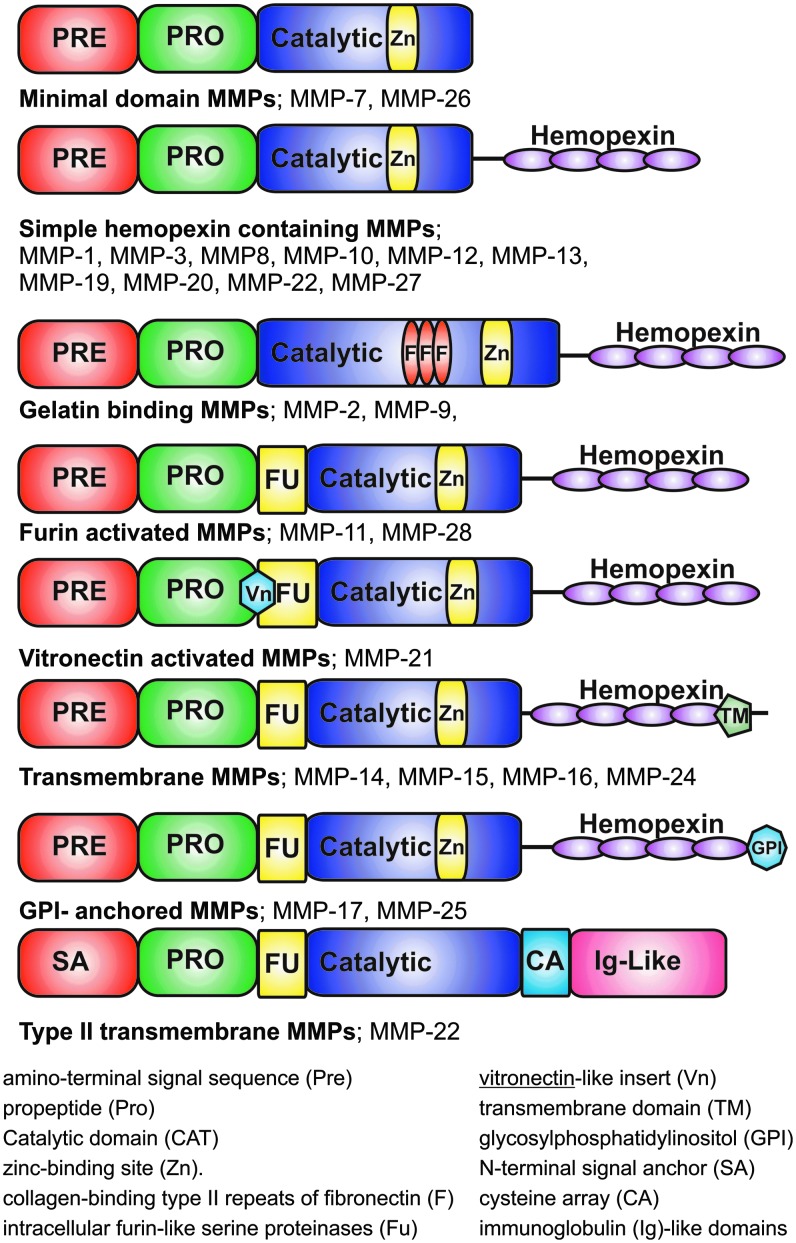
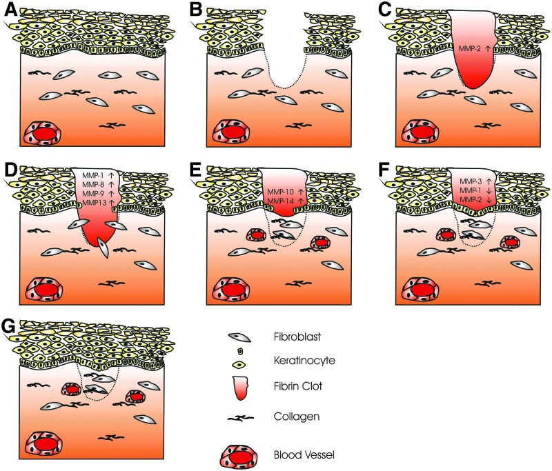
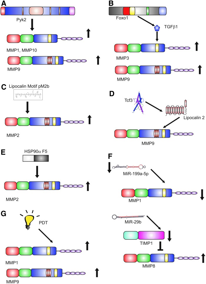
Significance: Matrix metalloproteinases (MMPs) are present in both acute and chronic wounds. They play a pivotal role, with their inhibitors, in regulating extracellular matrix degradation and deposition that is essential for wound reepithelialization. The excess protease activity can lead to a chronic nonhealing wound. The timed expression and activation of MMPs in response to wounding are vital for successful wound healing. MMPs are grouped into eight families and display extensive homology within these families. This homology leads in part to the initial failure of MMP inhibitors in clinical trials and the development of alternative methods for modulating the MMP activity. MMP-knockout mouse models display altered wound healing responses, but these are often subtle phenotypic changes indicating the overlapping MMP substrate specificity and inter-MMP compensation. Recent Advances: Recent research has identified several new MMP modulators, including photodynamic therapy, protease-absorbing dressing, microRNA regulation, signaling molecules, and peptides. Critical Issues: Wound healing requires the controlled activity of MMPs at all stages of the wound healing process. The loss of MMP regulation is a characteristic of chronic wounds and contributes to the failure to heal. Future Directions: Further research into how MMPs are regulated should allow the development of novel treatments for wound healing.


Figures

None
Matthew P. Caley, PhD
<b>Figure 1.</b>
Figure 1.
MMP family. MMPs can be divided into eight groups based on structural similarities and shared function. Minimal domain MMPs include MMP-7 and MMP-26. Simple hemopexin containing MMPs: MMP-1, MMP-3, MMP-8, MMP-10, MMP-12, MMP-13, MMP-19, MMP-20, MMP-22, and MMP-27. Gelatin binding MMPs: MMP-2 and MMP-9. Furin-activated MMPs: MMP-11 and MMP-28. Vitronectin-activated MMPs: MMP-21. Transmembrane MMPs: MMP-14, MMP-15, MMP-16, and MMP-24. GPI-anchored MMPs: MMP-17 and MMP-25. Type II transmembrane MMPs: MMP-22. MMP, matrix metalloproteinase.
<b>Figure 2.</b>
Figure 2.
Wound healing. Healthy skin consists of a stratified squamous epithelium, basement membrane, and dermis (A). The epithelium is made up of basal keratinocytes (proliferative) and stratified suprabasal keratinocytes (differentiating), which eventually lose their nuclear material. The dermis is made up of extracellular matrix, predominantly collagen, is populated by fibroblasts, and contains the blood vessels that supply the skin. (B) A full-thickness wound of the skin damaging both epidermis and dermis. (C) Inflammatory phase, the wound is filled with a fibrin clot sealing the wound, MMP-2 expression is increased. (D) Fibroblasts migrate into the wound area; using MMPs, they remodel the fibrin clot replacing it with new extracellular matrix. Epithelial cells upregulate MMP expression and migrate into the wound area (E). Failure to remodel the extracellular matrix due to increased MMP expression or inflammation is seen in chronic wounds and plays a part in their failure to heal. (F) Reepithelialization: epithelial cells migrate from the surrounding epithelium, proliferating and closing the wound. Expression of promigratory MMPs is decreased and tissue remodeling MMP expression is increased. (G) Wound maturation: epithelial cells proliferate and differentiate reforming the stratified squamous epithelium. Fibroblasts continue to remodel the underlying dermis over a period of several months.
<b>Figure 3.</b>
Figure 3.
Novel regulation of MMPs. Recent advances in the regulation of MMP expression and activity have used a variety of mechanisms. (A) Pyk2 has been shown to increase the expression of MMP-1, −9, and −10 in mouse models. (B) FOXO1, a member of the forkhead family of transcription factors, upregulates TGFβ1 leading to an increase in MMP-3 and MMP-9. (C) The 12 peptide fragment comprising the lipocalin conserved motif pM2b have been shown to increase MMP-2 in rat models of wounding. (D) Tcf3 increases secretion of lipocalin 2, which stabilizes MMP-9 increasing activity. (E) A 115 amino acid fragment (F5) of Hsp90α increases the secretion of MMP-2. (F) MiR, small hairpin loops of noncoding RNA, have been shown to regulate the MMP activity in wound models. (G) PDT has been shown to increase the expression of MMP-1 and MMP-9. MiR, microRNA; PDT, photodynamic therapy; Pyk2, proline-rich protein tyrosine kinase 2; Tcf3, transcription factor 3; TGF-β, transforming growth factor-β.

References

    1. Chen P, Parks WC. Role of matrix metalloproteinases in epithelial migration. J Cell Biochem 2009;108:1233–1243 - PMC - PubMed
    1. Zhang X, Nothnick WB. The role and regulation of the uterine matrix metalloproteinase system in menstruating and non-menstruating species. Front Biosci 2005;10:353–366 - PubMed
    1. Steffensen B, Hakkinen L, Larjava H. Proteolytic events of wound-healing—coordinated interactions among matrix metalloproteinases (MMPs), integrins, and extracellular matrix molecules. Crit Rev Oral Biol Med 2001;12:373–398 - PubMed
    1. Yan C, Boyd DD. Regulation of matrix metalloproteinase gene expression. J Cell Physiol 2007;211:19–26 - PubMed
    1. Toriseva M, Kahari VM. Proteinases in cutaneous wound healing. Cell Mol Life Sci 2009;66:203–224 - PMC - PubMed