. 2015 May;71(Pt 5):1102-11.
doi: 10.1107/S1399004715003521. Epub 2015 Apr 24.

Structures of the Middle East respiratory syndrome coronavirus 3C-like protease reveal insights into substrate specificity

Affiliations

Structures of the Middle East respiratory syndrome coronavirus 3C-like protease reveal insights into substrate specificity

Danielle Needle et al. Acta Crystallogr D Biol Crystallogr. 2015 May.

Abstract

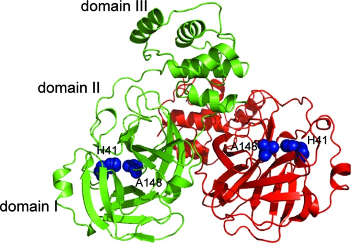
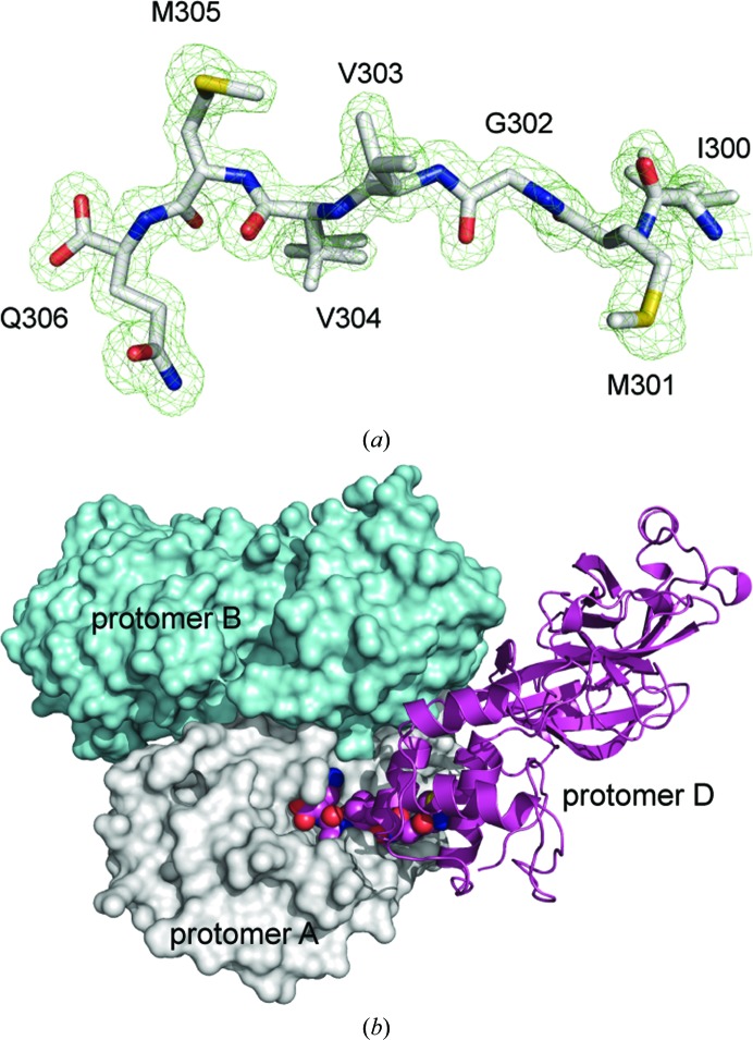
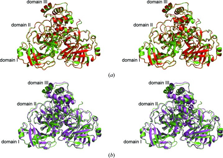
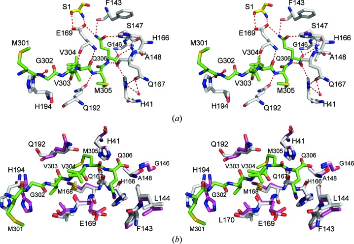
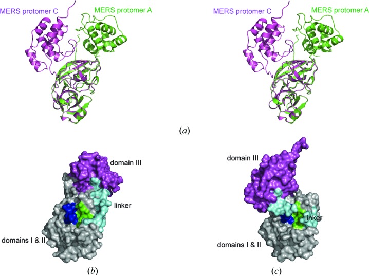
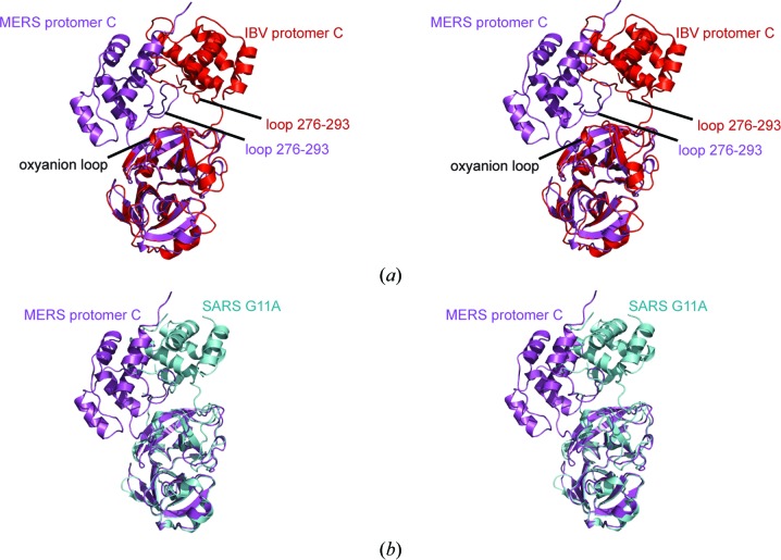
Middle East respiratory syndrome coronavirus (MERS-CoV) is a highly pathogenic virus that causes severe respiratory illness accompanied by multi-organ dysfunction, resulting in a case fatality rate of approximately 40%. As found in other coronaviruses, the majority of the positive-stranded RNA MERS-CoV genome is translated into two polyproteins, one created by a ribosomal frameshift, that are cleaved at three sites by a papain-like protease and at 11 sites by a 3C-like protease (3 CL(pro)). Since 3 CL(pro) is essential for viral replication, it is a leading candidate for therapeutic intervention. To accelerate the development of 3 CL(pro) inhibitors, three crystal structures of a catalytically inactive variant (C148A) of the MERS-CoV 3 CL(pro) enzyme were determined. The aim was to co-crystallize the inactive enzyme with a peptide substrate. Fortuitously, however, in two of the structures the C-terminus of one protomer is bound in the active site of a neighboring molecule, providing a snapshot of an enzyme-product complex. In the third structure, two of the three protomers in the asymmetric unit form a homodimer similar to that of SARS-CoV 3 CL(pro); however, the third protomer adopts a radically different conformation that is likely to correspond to a crystallographic monomer, indicative of substantial structural plasticity in the enzyme. The results presented here provide a foundation for the structure-based design of small-molecule inhibitors of the MERS-CoV 3 CL(pro) enzyme.

Keywords: 3CLpro; MERS-CoV; coronavirus; main protease.


Figures

Figure 1
Figure 1
The catalytically inactive MERS-CoV 3CLpro C148A homodimer as found in crystal form I. Protomer A is colored green and protomer B red. The residues forming the catalytic dyad are depicted as blue spheres.
Figure 2
Figure 2
(a) The C-terminal residues of protomer D (crystal form II), corresponding to the P6–P1 autoprocessed site of the mature enzyme fitted to the mF oDF c electron-density maps shown (contour level of 3.0σ, green; 1.55 Å resolution) after a round of refinement with the C-­terminal residues omitted from the model. (b) Illustration of the binding of the C-terminal tail (spheres) of protomer D (magenta ribbons) to the homodimer formed by protomer A (gray surface) and protomer B (cyan surface).
Figure 3
Figure 3
(a) Stereoview of the superimposed homodimers of MERS-CoV 3CLpro (crystal form II, green ribbons) and BatCoV-HKU4 (PDB entry 2yna, red ribbons). (b) Stereoview of the superimposed homodimers of MERS-CoV 3CLpro and SAR-CoV 3CLpro (PDB entry 1uk3, red ribbons; Yang et al., 2003 ▶).
Figure 4
Figure 4
Sequence alignment of CoV 3CLpro enzymes from MERS-CoV, SARS-CoV, Tylonycteris bat coronavirus HKU4, Human coronavirus HKU1, Human coronavirus OC43, Human coronavirus NL63 and Human coronavirus 229E. Sequences were aligned using T-Coffee (Notredame et al., 2000 ▶) and the figure was prepared with ESPript3 (Robert & Gouet, 2014 ▶). The residues forming the catalytic dyad are highlighted with asterisks.
Figure 5
Figure 5
(a) Stereoview of the hydrogen-bonding interactions (within 3.2 Å) between the C-terminal residues 301–306 of MERS-CoV 3CLpro protomer D (crystal form II, C atoms in green) and the active site of protomer A (C atoms in gray). Residue Ser1 (C atoms in yellow) is from protomer B of the homodimer. (b) Stereoview of the active-site residues from protomer A of the free enzyme form (crystal form I, C atoms in magenta) superimposed onto the active site of product-bound protomer A (crystal form II, C atoms in gray).
Figure 6
Figure 6
(a) Stereoview of the superimposed structures of MERS-CoV 3CLpro crystal form III protomer A (green ribbons) and protomer C (magenta ribbons). (b, c) Surface representations of protomer A (b) and protomer C (c) with domains I and II colored gray, the linker loop (residues 188–204) cyan, domain III magenta, the oxyanion loop (residues 143–148) blue and the S1 binding pocket green.
Figure 7
Figure 7
(a) Stereoview of the structure of MERS-CoV 3CLpro protomer C (crystal form III, magenta ribbons) superimposed on the structure of IBV-CoV 3CLpro protomer C (PDB entry 2q6d, red ribbons; Xue et al., 2008 ▶). (b) Stereoview of the structure of MERS-CoV 3CLpro protomer C (crystal form III, magenta ribbons) superimposed on the structure of the SARS-CoV 3CLpro G11A monomer (PDB entry 2pwx, cyan ribbons; Chen et al., 2008 ▶).

References

    1. Adams, P. D. et al. (2011). Methods, 55, 94–106.
    1. Adney, D. R., van Doremalen, N., Brown, V. R., Bushmaker, T., Scott, D., de Wit, E., Bowen, R. A. & Munster, V. J. (2014). Emerg. Infect. Dis. 20, 1999–2005. - PMC - PubMed
    1. Anand, K., Palm, G. J., Mesters, J. R., Siddell, S. G., Ziebuhr, J. & Hilgenfeld, R. (2002). EMBO J. 21, 3213–3224. - PMC - PubMed
    1. Anand, K., Ziebuhr, J., Wadhwani, P., Mesters, J. R. & Hilgenfeld, R. (2003). Science, 300, 1763–1767. - PubMed
    1. Artimo, P. et al. (2012). Nucleic Acids Res. 40, W597–W603. - PMC - PubMed

Publication types

MeSH terms