Host Response to the Lung Microbiome in Chronic Obstructive Pulmonary Disease
- PMID: 25945594
- PMCID: PMC4595667
- DOI: 10.1164/rccm.201502-0223OC
Host Response to the Lung Microbiome in Chronic Obstructive Pulmonary Disease
Abstract
Rationale: The relatively sparse but diverse microbiome in human lungs may become less diverse in chronic obstructive pulmonary disease (COPD). This article examines the relationship of this microbiome to emphysematous tissue destruction, number of terminal bronchioles, infiltrating inflammatory cells, and host gene expression.
Methods: Culture-independent pyrosequencing microbiome analysis was used to examine the V3-V5 regions of bacterial 16S ribosomal DNA in 40 samples of lung from 5 patients with COPD (Global Initiative for Chronic Obstructive Lung Disease [GOLD] stage 4) and 28 samples from 4 donors (controls). A second protocol based on the V1-V3 regions was used to verify the bacterial microbiome results. Within lung tissue samples the microbiome was compared with results of micro-computed tomography, infiltrating inflammatory cells measured by quantitative histology, and host gene expression.
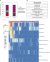
Measurements and main results: Ten operational taxonomic units (OTUs) was found sufficient to discriminate between control and GOLD stage 4 lung tissue, which included known pathogens such as Haemophilus influenzae. We also observed a decline in microbial diversity that was associated with emphysematous destruction, remodeling of the bronchiolar and alveolar tissue, and the infiltration of the tissue by CD4(+) T cells. Specific OTUs were also associated with neutrophils, eosinophils, and B-cell infiltration (P < 0.05). The expression profiles of 859 genes and 235 genes were associated with either enrichment or reductions of Firmicutes and Proteobacteria, respectively, at a false discovery rate cutoff of less than 0.1.
Conclusions: These results support the hypothesis that there is a host immune response to microorganisms within the lung microbiome that appears to contribute to the pathogenesis of COPD.
Keywords: COPD; bacteria; inflammation; microbiome.
Figures


Comment in
-
Outside In: Sequencing the Lung Microbiome.Am J Respir Crit Care Med. 2015 Aug 15;192(4):403-4. doi: 10.1164/rccm.201506-1138ED. Am J Respir Crit Care Med. 2015. PMID: 26278789 No abstract available.
-
The lung immune response to bacteria in chronic obstructive pulmonary disease.Am J Respir Crit Care Med. 2015 Oct 1;192(7):902-3. doi: 10.1164/rccm.201506-1117LE. Am J Respir Crit Care Med. 2015. PMID: 26426789 No abstract available.
-
Reply: the lung immune response to bacteria in chronic obstructive pulmonary disease.Am J Respir Crit Care Med. 2015 Oct 1;192(7):903-4. doi: 10.1164/rccm.201506-1257LE. Am J Respir Crit Care Med. 2015. PMID: 26426790 No abstract available.
References
-
- World Health Organization (WHO) Geneva, Switzerland: WHO Press; 2008. The global burden of disease: 2004 update.
-
- Baraldo S, Turato G, Saetta M. Pathophysiology of the small airways in chronic obstructive pulmonary disease. Respiration. 2012;84:89–97. - PubMed
-
- Di Stefano A, Turato G, Maestrelli P, Mapp CE, Ruggieri MP, Roggeri A, Boschetto P, Fabbri LM, Saetta M. Airflow limitation in chronic bronchitis is associated with T-lymphocyte and macrophage infiltration of the bronchial mucosa. Am J Respir Crit Care Med. 1996;153:629–632. - PubMed
-
- Hogg JC, Chu F, Utokaparch S, Woods R, Elliott WM, Buzatu L, Cherniack RM, Rogers RM, Sciurba FC, Coxson HO, et al. The nature of small-airway obstruction in chronic obstructive pulmonary disease. N Engl J Med. 2004;350:2645–2653. - PubMed
-
- Fletcher CM. Chronic bronchitis: its prevalence, nature, and pathogenesis. Am Rev Respir Dis. 1959;80:483–494. - PubMed
Publication types
MeSH terms
Grants and funding
LinkOut - more resources
Full Text Sources
Other Literature Sources
Medical
Research Materials

