

Modulation of cellular function through immune-activated exosomes
- PMID: 25945690
- PMCID: PMC4504252
- DOI: 10.1089/dna.2015.2884
Modulation of cellular function through immune-activated exosomes
Abstract
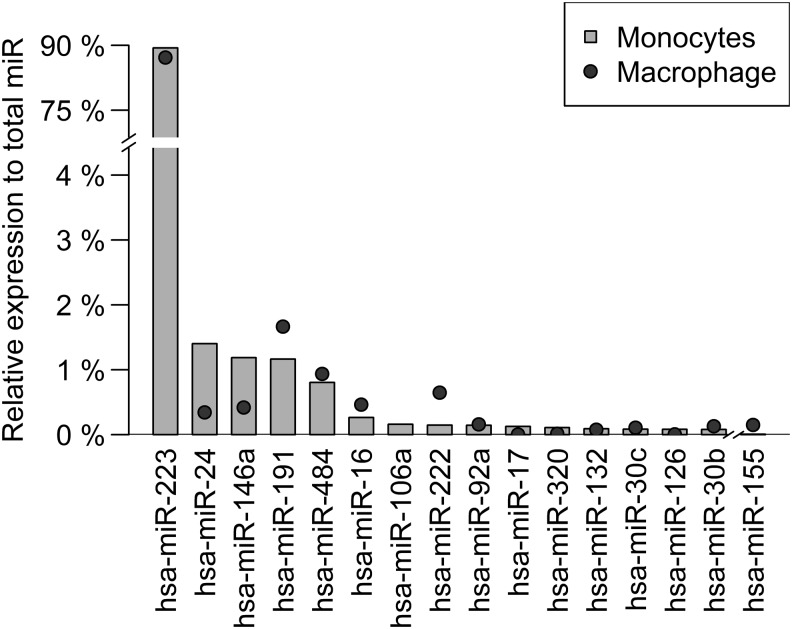
Extracellular vesicles classified as exosomes, microvesicles, or apoptotic bodies based on size are shed from most cells under normal as well as pathological conditions. They are released into the surrounding milieu, including plasma, urine, saliva, and tissues. Exosomes are highly enriched in microRNAs (miRs), which function in recipient cells by regulating posttranscriptional processing of targeted genes. Interaction of a miR with its mRNA target typically results in suppression of its gene expression. Peripheral inflammatory conditions can modulate miR expression in immune cells such as circulating monocytes that can influence their migration and differentiation. Changes within monocyte-derived macrophage miR expression can influence exosome content and further affect end-organ target cells.
Figures
Similar articles
-
Effects of exosomes from LPS-activated macrophages on adipocyte gene expression, differentiation, and insulin-dependent glucose uptake.J Physiol Biochem. 2018 Nov;74(4):559-568. doi: 10.1007/s13105-018-0622-4. Epub 2018 Mar 20. J Physiol Biochem. 2018. PMID: 29560554
-
Exosomes and microvesicles in normal physiology, pathophysiology, and renal diseases.Pediatr Nephrol. 2019 Jan;34(1):11-30. doi: 10.1007/s00467-017-3816-z. Epub 2017 Nov 27. Pediatr Nephrol. 2019. PMID: 29181712 Free PMC article. Review.
-
Hepatitis C virus direct-acting antivirals therapy impacts on extracellular vesicles microRNAs content and on their immunomodulating properties.Liver Int. 2018 Oct;38(10):1741-1750. doi: 10.1111/liv.13700. Epub 2018 Feb 24. Liver Int. 2018. PMID: 29359389
-
Extracellular Vesicles in Lung Disease.Chest. 2018 Jan;153(1):210-216. doi: 10.1016/j.chest.2017.06.026. Epub 2017 Jul 3. Chest. 2018. PMID: 28684288 Review.
-
Plasma exosome microRNAs are indicative of breast cancer.Breast Cancer Res. 2016 Sep 8;18(1):90. doi: 10.1186/s13058-016-0753-x. Breast Cancer Res. 2016. PMID: 27608715 Free PMC article.
Cited by
-
JC Polyomavirus Uses Extracellular Vesicles To Infect Target Cells.mBio. 2019 Apr 9;10(2):e00379-19. doi: 10.1128/mBio.00379-19. mBio. 2019. PMID: 30967463 Free PMC article.
-
Fifty Years of JC Polyomavirus: A Brief Overview and Remaining Questions.Viruses. 2020 Sep 1;12(9):969. doi: 10.3390/v12090969. Viruses. 2020. PMID: 32882975 Free PMC article. Review.
-
JC Virus infected choroid plexus epithelial cells produce extracellular vesicles that infect glial cells independently of the virus attachment receptor.PLoS Pathog. 2020 Mar 4;16(3):e1008371. doi: 10.1371/journal.ppat.1008371. eCollection 2020 Mar. PLoS Pathog. 2020. PMID: 32130281 Free PMC article.
-
Complications of COVID-19 on the Central Nervous System: Mechanisms and Potential Treatment for Easing Long COVID.Aging Dis. 2023 Oct 1;14(5):1492-1510. doi: 10.14336/AD.2023.0312. Aging Dis. 2023. PMID: 37163427 Free PMC article. Review.
-
Biomarkers of Activation and Inflammation to Track Disparity in Chronological and Physiological Age of People Living With HIV on Combination Antiretroviral Therapy.Front Immunol. 2020 Oct 9;11:583934. doi: 10.3389/fimmu.2020.583934. eCollection 2020. Front Immunol. 2020. PMID: 33162998 Free PMC article. Review.
References
-
- Biesmans S., Meert T.F., Bouwknecht J.A., Acton P.D., Davoodi N., De Haes P., Kuijlaars J., Langlois X., Matthews L.J., Ver Donck L., Hellings N., and Nuydens R. (2013). Systemic immune activation leads to neuroinflammation and sickness behavior in mice. Mediators Inflamm 2013, 271359. - PMC - PubMed
-
- Cheng L., Doecke J.D., Sharples R.A., Villemagne V.L., Fowler C.J., Rembach A., Martins R.N., Rowe C.C., Macaulay S.L., Masters C.L., and Hill A.F. (2014a). Prognostic serum miRNA biomarkers associated with Alzheimer's disease shows concordance with neuropsychological and neuroimaging assessment. Mol Psychiatry; doi:10.1038/mp.2014.127 - DOI - PubMed
Publication types
MeSH terms
Substances
Grants and funding
LinkOut - more resources
Full Text Sources
Other Literature Sources
