. 2015 May;71(Pt 5):553-9.
doi: 10.1107/S2053230X15000989. Epub 2015 Apr 21.

The X-ray structure of Plasmodium falciparum dihydroorotate dehydrogenase bound to a potent and selective N-phenylbenzamide inhibitor reveals novel binding-site interactions

Affiliations

The X-ray structure of Plasmodium falciparum dihydroorotate dehydrogenase bound to a potent and selective N-phenylbenzamide inhibitor reveals novel binding-site interactions

Xiaoyi Deng et al. Acta Crystallogr F Struct Biol Commun. 2015 May.

Abstract

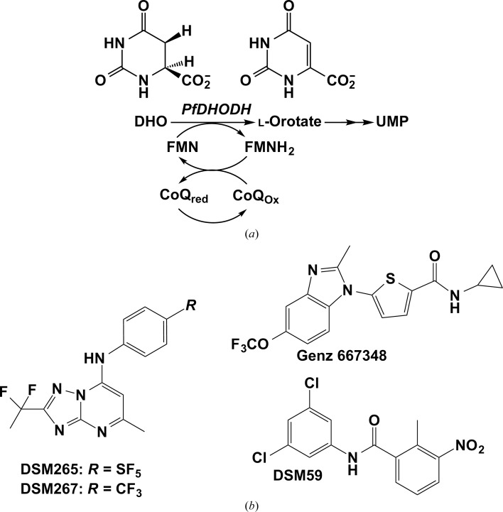
Plasmodium species are protozoan parasites that are the causative agent of malaria. Malaria is a devastating disease, and its treatment and control have been hampered by the propensity of the parasite to become drug-resistant. Dihydroorotate dehydrogenase (DHODH) has been identified as a promising new target for the development of antimalarial agents. Here, the X-ray structure of P. falciparum DHODH bound to a potent and selective N-phenylbenzamide-based inhibitor (DSM59) is described at 2.3 Å resolution. The structure elucidates novel binding-site interactions and shows how conformational flexibility of the enzyme leads to the ability to bind diverse chemical structures with high affinity. This information provides new insight into the design of high-affinity DHODH inhibitors for the treatment of malaria.

Keywords: DSM59; Plasmodium falciparum; dihydroorotate dehydrogenase.


Figures

Figure 1
Figure 1
(a) DHODH reaction scheme. (b) Chemical structures of PfDHODH inhibitors.
Figure 2
Figure 2
Electron-density map for PfDHODH–DSM59. The F oF c map calculated before the model was refined and prior to building in the ligand is contoured in green (+3σ) and red (−3σ). The 2F oF c map calculated after the model was refined contoured at 1σ is in blue. DSM59 is shown in pink and amino-acid residues are shown in yellow.
Figure 3
Figure 3
Structure of DSM59 bound to PfDHODH. (a) Ribbon diagram showing the DSM59 binding site on PfDHODH. DSM59 is displayed in pink, FMN in yellow and l-orotate in turquoise; protein C atoms are in turquoise, O atoms in red, N atoms in blue, phosphate in orange, S atoms in yellow and chloride in green. (b) Stereoview of the DSM59 binding site showing amino-acid residues within a 4 Å shell. DSM59 is displayed in pink, FMN, l-orotate and protein C atoms are in turquoise, O atoms in red, N atoms in blue, phosphate in orange, S atoms in yellow and chloride in green.
Figure 4
Figure 4
Inhibitor-binding site comparison of the binding modes of DSM59 (turquoise protein with DSM59 ligand in pink) and (a) DSM267 (purple) from the triazolopyrimidine chemical series and (b) Genz 667348 (purple) from the thiophene carboxamide chemical series. Selected amino-acid residues within 4 Å of the inhibitor-binding sites are displayed.

References

    1. Anthony, M. P., Burrows, J. N., Duparc, S., Moehrle, J. J. & Wells, T. N. (2012). Malar. J. 11, 316. - PMC - PubMed
    1. Ariey, F. et al. (2014). Nature (London), 505, 50–55. - PubMed
    1. Ashley, E. A. et al. (2014). N. Engl. J. Med. 371, 411–423. - PubMed
    1. Auffinger, P., Hays, F. A., Westhof, E. & Ho, P. S. (2004). Proc. Natl Acad. Sci. USA, 101, 16789–16794. - PMC - PubMed
    1. Baldwin, J., Michnoff, C. H., Malmquist, N. A., White, J., Roth, M. G., Rathod, P. K. & Phillips, M. A. (2005). J. Biol. Chem. 280, 21847–21853. - PubMed

Publication types

MeSH terms

Substances

Associated data