

Structural insights into thyroid hormone transport mechanisms of the L-type amino acid transporter 2
- PMID: 25945809
- PMCID: PMC5414734
- DOI: 10.1210/me.2015-1044
Structural insights into thyroid hormone transport mechanisms of the L-type amino acid transporter 2
Abstract
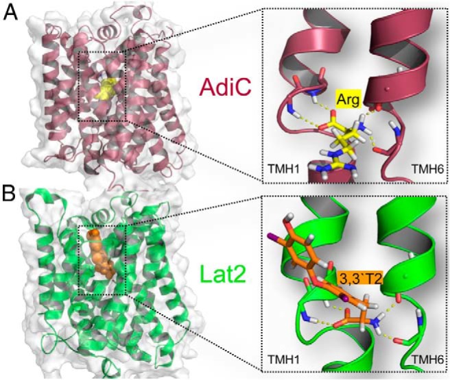
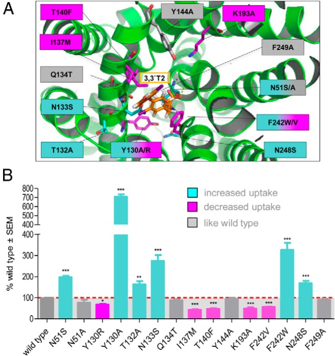
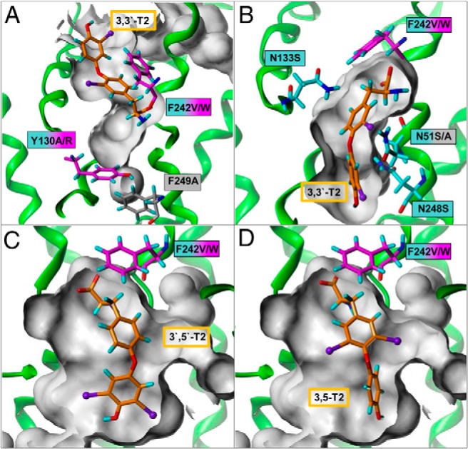
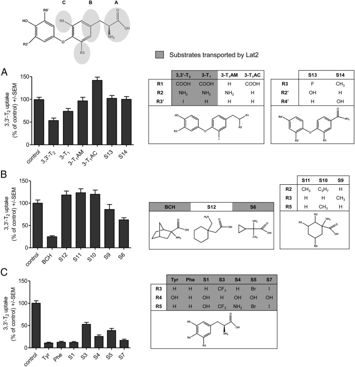
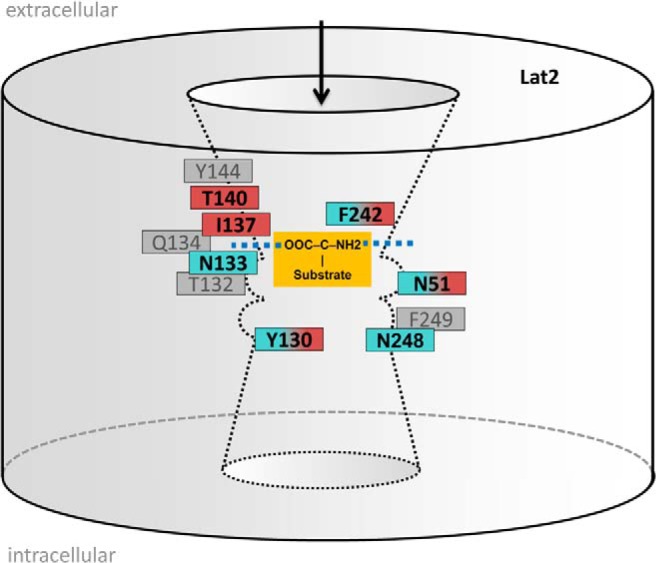
Thyroid hormones (THs) are transported across cell membranes by different transmembrane transporter proteins. In previous studies, we showed marked 3,3'-diiodothyronine (3,3'-T2) but moderate T3 uptake by the L-type amino acid transporter 2 (Lat2). We have now studied the structure-function relationships of this transporter and TH-like molecules. Our Lat2 homology model is based on 2 crystal structures of the homologous 12-transmembrane helix transporters arginine/agmatine antiporter and amino acid/polyamine/organocation transporter. Model-driven mutagenesis of residues lining an extracellular recognition site and a TH-traversing channel identified 9 sensitive residues. Using Xenopus laevis oocytes as expression system, we found that side chain shortening (N51S, N133S, N248S, and Y130A) expanded the channel and increased 3,3'-T2 transport. Side chain enlargements (T140F, Y130R, and I137M) decreased 3,3'-T2 uptake, indicating channel obstructions. The opposite results with mutations maintaining (F242W) or impairing (F242V) uptake suggest that F242 may have a gating function. Competitive inhibition studies of 14 TH-like compounds revealed that recognition by Lat2 requires amino and carboxylic acid groups. The size of the adjacent hydrophobic group is restricted. Bulky substituents in positions 3 and 5 of the tyrosine ring are allowed. The phenolic ring may be enlarged, provided that the whole molecule is flexible enough to fit into the distinctly shaped TH-traversing channel of Lat2. Taken together, the next Lat2 features were identified 1) TH recognition site; 2) TH-traversing channel in the center of Lat2; and 3) switch site that potentially facilitates intracellular substrate release. Together with identified substrate features, these data help to elucidate the molecular mechanisms and role of Lat2 in T2 transport.
Figures
Similar articles
-
Molecular features of the L-type amino acid transporter 2 determine different import and export profiles for thyroid hormones and amino acids.Mol Cell Endocrinol. 2017 Mar 5;443:163-174. doi: 10.1016/j.mce.2017.01.024. Epub 2017 Jan 18. Mol Cell Endocrinol. 2017. PMID: 28108384
-
Thyroid hormone transport across L-type amino acid transporters: What can molecular modelling tell us?Mol Cell Endocrinol. 2017 Dec 15;458:68-75. doi: 10.1016/j.mce.2017.03.018. Epub 2017 Mar 21. Mol Cell Endocrinol. 2017. PMID: 28341457 Review.
-
Involvement of the L-Type Amino Acid Transporter Lat2 in the Transport of 3,3'-Diiodothyronine across the Plasma Membrane.Eur Thyroid J. 2015 Sep;4(Suppl 1):42-50. doi: 10.1159/000381542. Epub 2015 May 28. Eur Thyroid J. 2015. PMID: 26601072 Free PMC article.
-
Transport of a neurotoxicant by molecular mimicry: the methylmercury-L-cysteine complex is a substrate for human L-type large neutral amino acid transporter (LAT) 1 and LAT2.Biochem J. 2002 Oct 1;367(Pt 1):239-46. doi: 10.1042/BJ20020841. Biochem J. 2002. PMID: 12117417 Free PMC article.
-
Pharmacokinetic role of L-type amino acid transporters LAT1 and LAT2.Eur J Pharm Sci. 2008 Oct 2;35(3):161-74. doi: 10.1016/j.ejps.2008.06.015. Epub 2008 Jul 5. Eur J Pharm Sci. 2008. PMID: 18656534 Review.
Cited by
-
Insights into the molecular basis for substrate binding and specificity of the wild-type L-arginine/agmatine antiporter AdiC.Proc Natl Acad Sci U S A. 2016 Sep 13;113(37):10358-63. doi: 10.1073/pnas.1605442113. Epub 2016 Aug 31. Proc Natl Acad Sci U S A. 2016. PMID: 27582465 Free PMC article.
-
Effects of Mutations and Ligands on the Thermostability of the l-Arginine/Agmatine Antiporter AdiC and Deduced Insights into Ligand-Binding of Human l-Type Amino Acid Transporters.Int J Mol Sci. 2018 Mar 20;19(3):918. doi: 10.3390/ijms19030918. Int J Mol Sci. 2018. PMID: 29558430 Free PMC article.
-
Reshaping the Binding Pocket of the Neurotransmitter:Solute Symporter (NSS) Family Transporter SLC6A14 (ATB0,+) Selectively Reduces Access for Cationic Amino Acids and Derivatives.Biomolecules. 2022 Oct 1;12(10):1404. doi: 10.3390/biom12101404. Biomolecules. 2022. PMID: 36291613 Free PMC article.
-
[Singleton and twin pregnancies of PKU patients - individual variability of phenylalanine tolerance: experience of a single treatment center (Preliminary report)].Dev Period Med. 2017;21(4):344-360. doi: 10.34763/devperiodmed.20172104.344360. Dev Period Med. 2017. PMID: 29291362 Free PMC article. Polish.
-
Function and Regulation of Acid Resistance Antiporters.J Membr Biol. 2019 Oct;252(4-5):465-481. doi: 10.1007/s00232-019-00073-6. Epub 2019 Jun 25. J Membr Biol. 2019. PMID: 31240358
References
-
- Rao GS, Eckel J, Rao ML, Breuer H. Uptake of thyroid hormone by isolated rat liver cells. Biochem Bioph Res Commun. 1976;73:98–104. - PubMed
-
- Abe T, Kakyo M, Sakagami H, et al. . Molecular characterization and tissue distribution of a new organic anion transporter subtype (oatp3) that transports thyroid hormones and taurocholate and comparison with oatp2. J Biol Chem. 1998;273:22395–22401. - PubMed
-
- Friesema EC, Ganguly S, Abdalla A, Manning Fox JE, Halestrap AP, Visser TJ. Identification of monocarboxylate transporter 8 as a specific thyroid hormone transporter. J Biol Chem. 2003;278:40128–40135. - PubMed
Publication types
MeSH terms
Substances
LinkOut - more resources
Full Text Sources
Other Literature Sources
Molecular Biology Databases
Miscellaneous
