. 2015 May 6;10(5):e0125946.
doi: 10.1371/journal.pone.0125946. eCollection 2015.

Structural Basis Underlying the Binding Preference of Human Galectins-1, -3 and -7 for Galβ1-3/4GlcNAc

Affiliations

Structural Basis Underlying the Binding Preference of Human Galectins-1, -3 and -7 for Galβ1-3/4GlcNAc

Tung-Ju Hsieh et al. PLoS One. .

Abstract

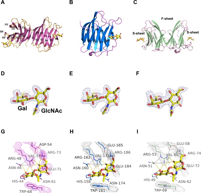
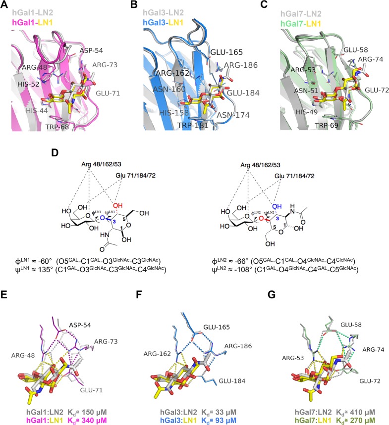
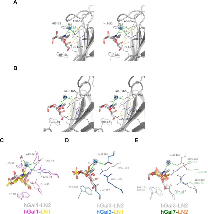
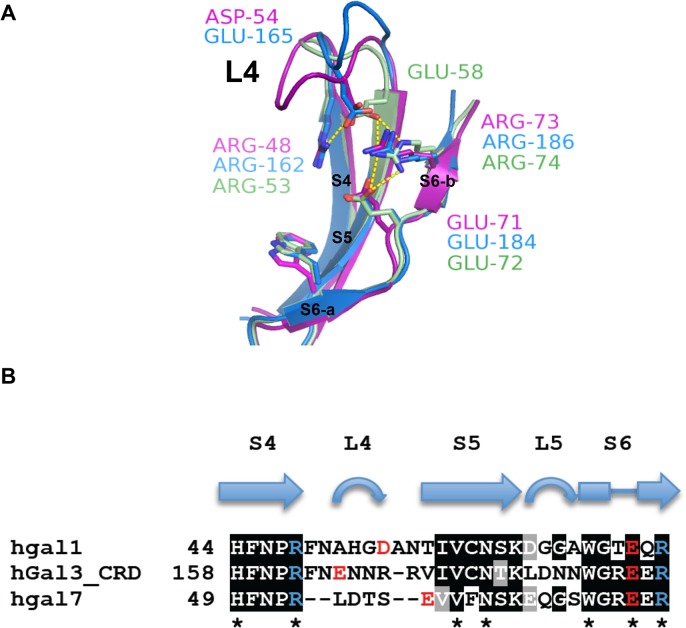
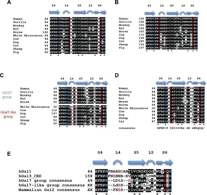
Galectins represent β-galactoside-binding proteins and are known to bind Galβ1-3/4GlcNAc disaccharides (abbreviated as LN1 and LN2, respectively). Despite high sequence and structural homology shared by the carbohydrate recognition domain (CRD) of all galectin members, how each galectin displays different sugar-binding specificity still remains ambiguous. Herein we provided the first structural evidence of human galectins-1, 3-CRD and 7 in complex with LN1. Galectins-1 and 3 were shown to have higher affinity for LN2 than for LN1, while galectin-7 displayed the reversed specificity. In comparison with the previous LN2-complexed structures, the results indicated that the average glycosidic torsion angle of galectin-bound LN1 (ψ(LN1) ≈ 135°) was significantly differed from that of galectin-bound LN2 (ψ(LN2 )≈ -108°), i.e. the GlcNAc moiety adopted a different orientation to maintain essential interactions. Furthermore, we also identified an Arg-Asp/Glu-Glu-Arg salt-bridge network and the corresponding loop (to position the second Asp/Glu residue) critical for the LN1/2-binding preference.


Conflict of interest statement

Competing Interests: The authors have declared that no competing interests exist.

Figures

Fig 1
Fig 1. Structural overview of hGal1, 3-CRD and 7 in complex with LN1.
(A-C) Ribbon representations of three LN1-hGal complexes where LN1 (shown in yellow stick model) is bound to hGal1 (A), hGal3-CRD (B) and hGal7 (C). Numbering of the β-strands of S-sheet (S1-S6) and F-sheet (F1-F5) is also shown as indicated. (D-F) F o -F c omit electron density map of LN1 (contoured at 2.5σ) bound to hGal1 (D), hGal3-CRD (E) and hGal7 (F). To make it clear, carbons 1, 3 and 5 of Gal and GlcNAc are labeled. (G-I) β-galactoside-recognition site of hGal1-LN1 (G), hGal3-CRD-LN1 (H) and hGal7-LN1 (I) complexes. Residues involved in LN1 recognition are highlighted with 2F o -F c electron density (contoured at 1σ). Polar interactions among galectin residues and within galectin/LN1 complex are shown as gray and yellow dash lines, respectively.
Fig 2
Fig 2. Pairwise comparison of the β-galactoside-recognition site in the LN1-hGal1/3-CRD/7 and LN2-hGal1/3-CRD/7 complexes.
(A-C) Structures of hGal1 (PDB ID: 1W6P), hGal3-CRD (1KJL) and hGal7 (5GAL) in complex with LN2 (all shown in gray) were superimposed, respectively, with the LN1 (in yellow)-containing structures of hGal1 (pink), hGal3-CRD (cyan) and hGal7 (green). (D) Diagrams delineate the different interaction geometries of LN1 (left) and LN2 (right) with respect to hGal1/3-CRD/7. Definition and values of the glycosidic torsion angles (ϕ and ψ) for LN1 and LN2 molecules are also listed. (E-G) Close-up view of the unique salt bridge networks in hGal1 (E), hGal3-CRD (F) and hGal7 (G), with a superposition of their LN2 and LN1-complex structures. Polar interactions among LN2-complexed structures are shown in gray dash lines while the interactions among amino acid residues in LN1-loaded structures are indicated by colored dash lines according to hGal1 (pink), 3-CRD (blue) and 7 (green), respectively, and all interactions related to galectin/LN1 complexes formation are colored in yellow.
Fig 3
Fig 3. Water-mediated interactions of hGal1 and hGal3 with the LN2 molecules.
(A and B) Stereoview of LN2 molecule bound in the carbohydrate-recognition site of hGal1 (PDB ID: 1W6P) and hGal3-CRD (1KJL), respectively. LN2 ligand is depicted as gray stick model. The water (blue sphere) is coordinated by the H-bonds (green dashes) from N2 atom of LN2 and amino acid residues of galectins. 2F o -F c omit electron density (gray mesh) of the water molecules are highlighted and contoured at 1σ. Unique salt bridge network of hGal1 and hGal3-CRD are indicated as yellow dashes. (C and D) Structural superposition of LN1 and LN2 complex structures from hGal1 (C) and hGal3-CRD (D). The C5-hydroxyl group of LN1 (yellow sticks) in hGal1 and hGal3-CRD complexes makes close contact with the coordinated water in LN2-hGal1 andLN2-hGal3-CRD complex with a distance of 2.2 and 2.1 Å, respectively. (E) Structure of hGal7 (shown in color green) in complex with LN2 (orange) is superimposed with LN2-hGal3-CRD complex structure (all in gray). Most LN2-contacting residues in hGal7 (such as Arg53hGal7, Trp69hGal7, Glu72hGal7 and Arg74hGal7) are well superimposed with those of hGal3, except the Glu58hGal7 residue. Location of Glu58hGal7 is distinctive from Glu165hGal3/ Asp54hGal1 and distance between Glu58hGal7 and N2 atom of LN2 is too far for them to coordinate a water molecule in between.
Fig 4
Fig 4. Structural comparisons among hGal1, 3-CRD and 7.
(A) S4-S6 β-strands of hGal1 (pink), 3-CRD (blue) and 7 (green) are superimposed. The unique salt bridge network of hGal1 (R48-D54-E71-R73), 3-CRD (R162-E165-E184-R186) and 7 (R53-E58-E72-R74) are shown in stick models. (B) Structure-based sequence alignment of S4-S6 β-strands of hGal1, 3-CRD and 7. Secondary structures were designated according to the resolved x-ray structures. The highly conserved LNs-interacting residues among hGal1, 3-CRD and 7 are indicated by asterisks. Residues involved in unique salt bridge network of hGal1, 3-CRD and 7 are colored in either red (Glu/Asp) or blue (Arg).
Fig 5
Fig 5. Conservation of the L4 regions among mammalian galectins.
Structure-based sequence alignment of S4-S6 β-strands of mammalian galectin-1s (A), galectin-3s (B), galectin-7s (C) and galectin-2s (D). According to the variation at corresponding position of Glu58hGal7, mammalian galectin-7s are further divided into two subgroups, the hGal7 group and hGal7-llike group. Sequence comparison of L4 regions of human Galectin-1, 3 and mammalian Galectin-2, 7 (E). Invariant LNs-contacting residues of galectins are indicated by asterisks. Residues involved in unique salt-bridge network are colored according to negative (red) or positive (blue) charged properties. The sequences of mammalian galectins were selected from human (Homo sapiens: galectin-1 [NP_002296.1], galectin-2 [NP_006489.1], galectin-3 [BAA22164.1] and galectin-7 [NP_002298.1]), Gorilla (Gorilla gorilla gorilla: galectin-1 [XP_004063482.1], galectin-2 [XP_004063485.1], galectin-3 [XP_004055252.1] and galectin-7 [XP_004060726.1]), Monkey (Macaca mulatta: galectin-1 [NP_001162098.1], galectin-2 [XP_001087063.1], galectin-3 [NP_001253292.1] and galectin-7 [NP_001083444.1]), Rat (Rattus norvegicus: galectin-1 [NP_063969.1], galectin-2 [NP_598283.1], galectin-3 [NP_114020.1] and galectin-7 [NP_072104.2]), Horse (Equus caballus: galectin-1 [XP_001501082.2], galectin-2 [XP_001499566.2], galectin-3 [XP_005605252.1] and galectin-7 [XP_001496714.2]), Rhinoceros (Ceratotherium simum simum: galectin-1 [XP_004418181.1], galectin-2 [XP_004418176.1] and galectin-7 [XP_004441551.1]), Cow (Bos taurus: galectin-1 [NP_786976.1], galectin-2 [NP_001244020.1], galectin-3 [NP_001095811.1] and galectin-7 [XP_002695023.2]), Dog (Canis lupus familiaris: galectin-1 [ADR80617.1], galectin-2 [NP_001271396.1], galectin-3 [NP_001183972.1] and galectin-7 [NP_001183972.1]), Cat (Felis catus: galectin-1 [XP_003989294.1], galectin-2 [XP_006934157.1], galectin-3 [XP_003987704.1] and galectin-7 [XP_003987704.1]), Sheep (Ovis aries: galectin-1 [AAT38511.1], galectin-2 [XP_004007664.1], galectin-3 [XP_004010713.1] and galectin-7 [XP_004010713.1]), Pig (Sus scrofa: galectin-1 [NP_001001867.1], galectin-3 [NP_001090970.1] and galectin-7 [NP_001136315.1]) and Mouse (Mus Musculus: galectin-2 [NP_079898.2]).

Similar articles

Cited by

References

    1. Barondes SH, Castronovo V, Cooper DN, Cummings RD, Drickamer K, Feizi T, et al. Galectins: a family of animal beta-galactoside-binding lectins. Cell. 1994. February 25;76(4):597–8. - PubMed
    1. Leffler H, Carlsson S, Hedlund M, Qian Y, Poirier F. Introduction to galectins. Glycoconjugate journal. 2004;19(7–9):433–40. - PubMed
    1. Rabinovich GA, Toscano MA, Jackson SS, Vasta GR. Functions of cell surface galectin-glycoprotein lattices. Curr Opin Struct Biol. 2007. October;17(5):513–20. - PMC - PubMed
    1. Yang RY, Liu FT. Galectins in cell growth and apoptosis. Cell Mol Life Sci. 2003. February;60(2):267–76. - PMC - PubMed
    1. Liu FT, Rabinovich GA. Galectins as modulators of tumour progression. Nat Rev Cancer. 2005. January;5(1):29–41. - PubMed

Publication types