Review
. 2015 May 4;5(2):702-23.
doi: 10.3390/biom5020702.

The Role of Reactive Oxygen Species (ROS) in the Formation of Extracellular Traps (ETs) in Humans

Affiliations
Review

The Role of Reactive Oxygen Species (ROS) in the Formation of Extracellular Traps (ETs) in Humans

Walter Stoiber et al. Biomolecules. .

Abstract

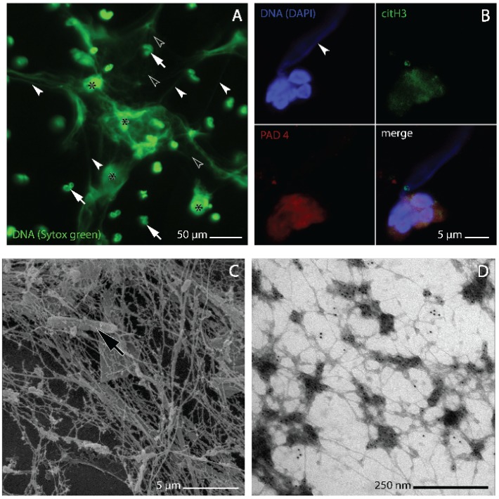
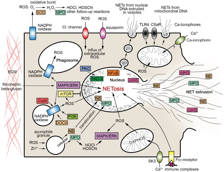
Extracellular traps (ETs) are reticulate structures of extracellular DNA associated with antimicrobial molecules. Their formation by phagocytes (mainly by neutrophils: NETs) has been identified as an essential element of vertebrate innate immune defense. However, as ETs are also toxic to host cells and potent triggers of autoimmunity, their role between pathogen defense and human pathogenesis is ambiguous, and they contribute to a variety of acute and chronic inflammatory diseases. Since the discovery of ET formation (ETosis) a decade ago, evidence has accumulated that most reaction cascades leading to ET release involve ROS. An important new facet was added when it became apparent that ETosis might be directly linked to, or be a variant of, the autophagy cell death pathway. The present review analyzes the evidence to date on the interplay between ROS, autophagy and ETosis, and highlights and discusses several further aspects of the ROS-ET relationship that are incompletely understood. These aspects include the role of NADPH oxidase-derived ROS, the molecular requirements of NADPH oxidase-dependent ETosis, the roles of NADPH oxidase subtypes, extracellular ROS and of ROS from sources other than NADPH oxidase, and the present evidence for ROS-independent ETosis. We conclude that ROS interact with ETosis in a multidimensional manner, with influence on whether ETosis shows beneficial or detrimental effects.


Figures

Figure 1
Figure 1
Morphology of NETs and NET-forming human neutrophils as analyzed by confocal laser microscopy (A, B), and scanning and transmission electron microscopy (C and D, respectively). (A) NETs generated in vitro from human neutrophils isolated from whole venous blood using a standard gradient separation medium containing sodium metrizoate and Dextran 500 [34]. NETosis induction by stimulation with 1 µM fMLP followed the procedure described by [35]. DNA was stained with sytox® green. At light microscopic resolution, NETs appear as irregular cloudy structures in which dense clusters of brightly stained extracellular DNA (asterisks) merge with more faintly stained dilated areas in which the DNA is more thinly spread and forms a meshwork of threads (white arrowheads). Lobulated nuclei of non-NET forming neutrophils (white arrows) are found within the meshwork as well as outside of it, some being slightly out of focus; (B) elongated plume of NET-DNA (arrowhead) protruding from one of two attached NET-forming neutrophils from the sputum of a patient with chronic obstructive pulmonary disease (COPD). The cells are immunostained for peptidyl arginine deiminase 4 (PAD4, red) and citrullinated histone 3 (citH3, green), DNA is stained with 4',6-diamidino-2-phenylindole (DAPI, blue). Overlapping PAD4 and citH3 staining at nuclear and cytoplasmic sites is characteristic of NET-forming neutrophils (cf. [9,36]) and conforms with the observation of [37] that histone H3 deimination by PAD4 is not entirely confined to the nucleus; (C) bacterium (arrow) entangled in NETs from the sputum of a COPD patient; (D) on-grid preparation of in vitro generated NETs (procedures as described for A above) immunogold stained for the enzyme neutrophil elastase, one of the key protein components of NETs.
Figure 2
Figure 2
Scheme summarizing pathways of interaction between ROS and NET formation as addressed in the text. Arrows indicate directions of effects. Note that only some of the processes shown can co-occur. CatS cathepsin S, citH3 citrullinated histone H3, C5aR complement component 5a receptor, DOCK dedicator of cytokinesis proteins, ERK extracellular signal-regulated kinases, HOCl hypochlorous acid, HOCSN hypothiocyanous acid, MASPK mitogen-activated protein kinases, MPO myeloperoxidase, mTOR mammalian target of rapamycin, NE neutrophil elastase, NFκB nuclear factor kappa-light-chain-enhancer of activated B cells, OXPHOS oxidative phosphorylation, PAD4 peptidylarginine deiminase 4, PI3K phosphoinositide-3-kinase, PKC protein kinase C, SK3 small conductance calcium-activated potassium channel 3, SOD superoxide dismutase, TLR4 toll-like receptor 4.

References

    1. Von Köckritz-Blickwede M., Nizet V. Innate immunity turned inside-out: Antimicrobial defense by phagocyte extracellular traps. J. Mol. Med. 2009;87:775–783. - PMC - PubMed
    1. Remijsen Q., Kuijpers T., Wirawan E., Lippens S., Vandenabeele P., Berghe T.V. Dying for a cause: NETosis, mechanisms behind an antimicrobial cell death modality. Cell Death Differ. 2011;18:581–588. doi: 10.1038/cdd.2011.1. - DOI - PMC - PubMed
    1. Guimarães-Costa A.B., Nascimento M.T.C., Wardini A.B., Pinto-da-Silva L.H., Saraiva E.M. ETosis: A microbicidal mechanism beyond Cell Death. J. Parasitol. Res. 2012 doi: 10.1155/2012/929743. - DOI - PMC - PubMed
    1. Goldmann O., Medina E. The expanding world of extracellular traps: Not only neutrophils but much more. Front. Immunol. 2013 doi: 10.3389/fimmu.2012.00420. - DOI - PMC - PubMed
    1. Brinkmann V., Reichard U., Goosmann C., Fauler B., Uhlemann Y., Weiss D.S., Weinrauch Y., Zychlinsky A. Neutrophil extracellular traps kill bacteria. Science. 2004;303:1532–1535. doi: 10.1126/science.1092385. - DOI - PubMed