. 2016 Mar;75(3):1301-11.
doi: 10.1002/mrm.25731. Epub 2015 May 6.

High temporal resolution dynamic MRI and arterial input function for assessment of GFR in pediatric subjects

Affiliations

High temporal resolution dynamic MRI and arterial input function for assessment of GFR in pediatric subjects

Umit Yoruk et al. Magn Reson Med. 2016 Mar.

Abstract

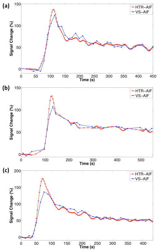
Purpose: To introduce a respiratory-gated high-spatiotemporal-resolution dynamic-contrast-enhanced MRI technique and a high-temporal-resolution aortic input function (HTR-AIF) estimation method for glomerular filtration rate (GFR) assessment in children.

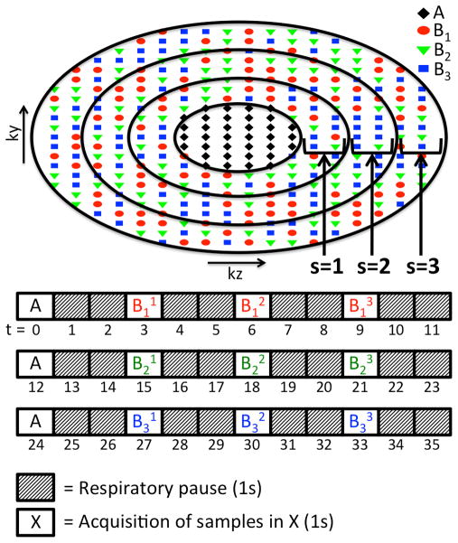
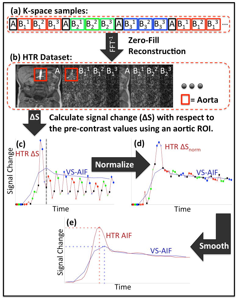
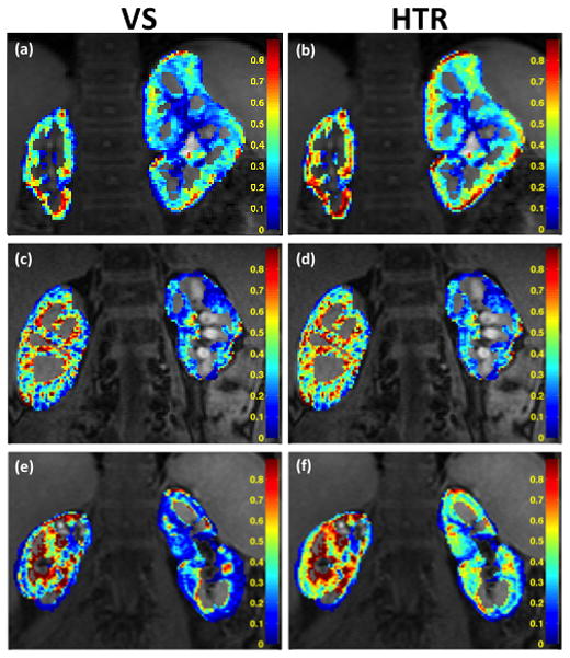
Methods: A high-spatiotemporal-resolution DCE-MRI method with view-shared reconstruction was modified to incorporate respiratory gating, and an AIF estimation method that uses a fraction of the k-space data from each respiratory period was developed (HTR-AIF). The method was validated using realistic digital phantom simulations and demonstrated on clinical subjects. The GFR estimates using HTR-AIF were compared with estimates obtained by using an AIF derived directly from the view-shared images.

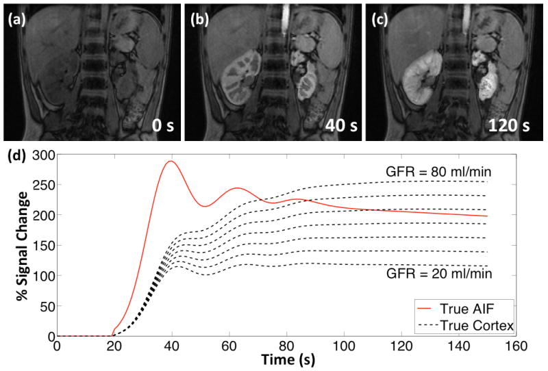
Results: Digital phantom simulations showed that using the HTR-AIF technique gives more accurate AIF estimates (RMSE = 0.0932) compared with the existing estimation method (RMSE = 0.2059) that used view-sharing (VS). For simulated GFR > 27 mL/min, GFR estimation error was between 32% and 17% using view-shared AIF, whereas estimation error was less than 10% using HTR-AIF. In all clinical subjects, the HTR-AIF method resulted in higher GFR estimations than the view-shared method.

Conclusion: The HTR-AIF method improves the accuracy of both the AIF and GFR estimates derived from the respiratory-gated acquisitions, and makes GFR estimation feasible in free-breathing pediatric subjects.

Keywords: arterial input function estimation; dynamic contrast enhancement; glomerular filtration rate estimation; high spatiotemporal resolution dynamic imaging; urography.


Figures

FIG. 1
FIG. 1
DISCO sampling pattern (fA = 0.1, M = 3, S = 3) and timing of acquisition assuming a respiratory rate of 20 breaths/min with end-expiratory phase lasting 1s at each cycle. Each sub-region of B is represented with Bms where the subscript “m” denotes the pseudo-randomly subsampled regions (m = 1 to M), which are also color coded in the figure, and superscript “s” denotes the annular sub-regions (s = 1 to S).
FIG. 2
FIG. 2
Flowchart of HTR-AIF analysis: K-space samples from the same respiratory cycle are grouped together (a). Volumetric images are reconstructed from each group of samples to form the HTR dataset (b). Signal change within the aorta is calculated (c). The dashed line marks the peak time of the VS-AIF curve (blue), which marks the start of the tail section. The signal change curve (red) is normalized using the tail section of the VS-AIF curve as a reference (d). The data points corresponding to A regions in (c) and (d) are marked with black markers as a reference to demonstrate the scaling differences between different groups. The final HTR-AIF estimate is obtained by applying local regression to the normalized signal change (e). Depending on the timing and the temporal dynamics of the true AIF, the HTR-AIF and VS-AIF estimates may give different peak heights and times.
FIG. 3
FIG. 3
Digital phantom at (a) pre-contrast, (b) cortical enhancement and (c) medullary enhancement phases. (d) The simulated AIF and cortical enhancement curves (20 mL/min to 80 mL/min single kidney GFR).
FIG. 4
FIG. 4
Digital phantom simulation results: (a) The HTR-AIF and VS-AIF estimates are compared to the true AIF of the phantom (only one of the 12 curves shown), (b) median GFR estimation errors obtained from the quantitative analysis of the simulated phantoms using HTR-AIF and VS-AIF methods.
FIG. 5
FIG. 5
Selected slices from pre-contrast (a) and post-contrast (b, d, e) images of the 5-year-old patient show different stages of contrast enhancement in kidneys. Maximum intensity projection (c) reveals arteries during the arterial enhancement phase. Panel (f) shows the segmentation map of the kidneys, which is needed for quantitative analysis.
FIG. 6
FIG. 6
HTR-AIF and VS-AIF estimates for the 3 clinical cases (a) 5 year old (b) 9 year old (c) 17 year old.
FIG. 7
FIG. 7
GFR Maps of 5-year-old (a–b), 9-year-old (c–d) and 17-year-old (e–f) subjects calculated using VS-AIF (left column) and HTR-AIF (right column). The reported values are GFR per mL of tissue (k21).

References

    1. Wong CJ, Moxey-Mims M, Jerry-Fluker J, Warady BA, Furth SL. CKiD (CKD in Children) Prospective Cohort Study: A Review of Current Findings. American Journal of Kidney Diseases. 2012;60(6):1002–1011. - PMC - PubMed
    1. Warady BA, Chadha V. Chronic kidney disease in children: the global perspective. Pediatric Nephrology. 2007;22(12):1999–2009. - PMC - PubMed
    1. Sigmund G, Stoever B, Zimmerhackl LB, Frankenschmidt A, Nitzsche E, Leititis JU, Struwe FE, Hennig J. RARE-MR-urography in the diagnosis of upper urinary tract abnormalities in children. Pediatric radiology. 1991;21(6):416–420. - PubMed
    1. Regan F, Bohlman ME, Khazan R, Rodriguez R, Schultze-Haakh H. MR urography using HASTE imaging in the assessment of ureteric obstruction. AJR American journal of roentgenology. 1996;167(5):1115–1120. - PubMed
    1. Aerts P, Van Hoe L, Bosmans H, Oyen R, Marchal G, Baert AL. Breath-hold MR urography using the HASTE technique. AJR American journal of roentgenology. 1996;166(3):543–545. - PubMed

Publication types