. 2015;11(5):785-95.
doi: 10.1080/15548627.2015.1037061.

SYK regulates macrophage MHC-II expression via activation of autophagy in response to oxidized LDL

Affiliations

SYK regulates macrophage MHC-II expression via activation of autophagy in response to oxidized LDL

Soo-Ho Choi et al. Autophagy. 2015.

Abstract

Adaptive immunity, which plays an important role in the development of atherosclerosis, is mediated by major histocompatibility complex (MHC)-dependent antigen presentation. In atherosclerotic lesions, macrophages constitute an important class of antigen-presenting cells that activate adaptive immune responses to oxidized low-density lipoprotein (OxLDL). It has been reported that autophagy regulates adaptive immune responses by enhancing antigen presentation to MHC class II (MHC-II). In a previous study, we have demonstrated that SYK (spleen tyrosine kinase) regulates generation of reactive oxygen species (ROS) and activation of MAPK8/JNK1 in macrophages. Because ROS and MAPK8 are known to regulate autophagy, in this study we investigated the role of SYK in autophagy, MHC-II expression and adaptive immune response to OxLDL. We demonstrate that OxLDL induces autophagosome formation, MHC-II expression, and phosphorylation of SYK in macrophages. Gene knockout and pharmacological inhibitors of NOX2 and MAPK8 reduced OxLDL-induced autophagy. Using bone marrow-derived macrophages isolated from wild-type and myeloid-specific SYK knockout mice, we demonstrate that SYK regulates OxLDL-induced ROS generation, MAPK8 activation, BECN1-BCL2 dissociation, autophagosome formation and presentation of OxLDL-derived antigens to CD4(+) T cells. ldlr(-/-) syk(-/-) mice fed a high-fat diet produced lower levels of IgG to malondialdehyde (MDA)-LDL, malondialdehyde-acetaldehyde (MAA)-LDL, and OxLDL compared to ldlr(-/-) mice. These results provide new insights into the mechanisms by which SYK regulates MHC-II expression via autophagy in macrophages and may contribute to regulation of adaptive immune responses in atherosclerosis.

Keywords: 3MA, 3-methyladenine; APCs, antigen-presenting cells; BCR, B cell receptor; BMDM, bone marrow-derived macrophage; Baf, bafilomycin A1; DPI, diphenyleneiodonium; FCGR, Fc fragment of IgG; GFP, green fluorescent protein; HFD, high-fat diet; IL2, interleukin 2; ITAM, immunoreceptor tyrosine-based activation motif; IgG, immunoglobulin G; IgM, immunoglobulin M; LPS, lipopolysaccharide; MAA-LDL, malondialdehyde-acetaldehyde modified low density lipoprotein; MAP1LC3/LC3, microtubule-associated protein 1 light chain 3; MAPK, mitogen-activated protein kinase; MDA-LDL, malondialdehyde modified low density lipoprotein; MHC-II; MHC-II, major histocompatibility complex class II; NOX, NAPDH oxidase; OSE, oxidation specific epitopes; OxLDL; OxLDL, oxidized low density lipoprotein; PBS, phosphate-buffered saline; PIC, piceatannol; ROS; ROS, reactive oxygen species; SYK; SYK, spleen tyrosine kinase; TCR, T cell receptor; TLR4, toll-like receptor 4; TNF, tumor necrosis factor; autophagy; low affinity, receptor; mmLDL, minimally modified low density lipoprotein; oxidation-specific antibodies.


Figures

Figure 1.
Figure 1.
OxLDL upregulates surface expression of MHC-II on macrophages, both in vitro and in vivo. (A) BMDM isolated from C57BL/6 mice were incubated with PBS or 25 μg/ml OxLDL for 18 h and then analyzed for MHC-II expression by FACS. (B) Quantification of the results presented in panel A. (C) C57BL/6 mice were intraperitoneally injected with 0.2 ml PBS or 0.2 ml of 500 μg/ml OxLDL. After 24 h, peritoneal cells were isolated and the MHC-II expression on F4/80-positive macrophages was analyzed by FACS. (D) Quantification of the results presented in panel C. Mean± SE ; n = 3 −4. *, P < 0.05; **, P < 0.005.
Figure 2.
Figure 2.
OxLDL induces autophagy in macrophages in vitro and in vivo. (A) RAW264.7 cells were incubated with 25 μg/ml of OxLDL for 18 h. LC3 and GAPDH were detected by immunoblot. (B) RAW264.7 cells stably expressing GFP-LC3B were incubated with 25 μg/ml OxLDL for 18 h. The pattern of GFP-LC3B localization was visualized by deconvolution microscopy. Hoechst 33358 was used to visualize nuclei (blue). (C and D) BMDM isolated from C57BL/6 mice were pretreated with or without 100 nM Baf for 1 h and then incubated with 25 μg/ml OxLDL for 18 h. Cell lysates were immunoblotted with the indicated antibodies, and the band densities were quantified. (E) BMDM incubated with OxLDL as in panel C were stained with anti-LC3 and anti-SQSTM1 antibodies and Hoechst 33358. (F to I) C57BL/6 mice were intraperitoneally injected with 0.2 ml of PBS or 0.2 ml of 500 μg/ml OxLDL. After 24 h, peritoneal cells were isolated and plated for 2 h. (F and G) Cell lysates were immunoblotted with the indicated antibodies and band intensities were quantified. (H and I) Cells were stained with an anti-LC3 antibody and Hoechst 33358 and the numbers of LC3 puncta per cell were counted. Mean ± SE ; n = 3 −4. *, P < 0.05; **, P < 0.005; ***, P < 0.0005. Scale bar: 10 μm.
Figure 3.
Figure 3.
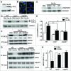
Involvement of SYK, NOX2, and MAPK8 in OxLDL-induced autophagy. (A) BMDM isolated from C57BL/6 mice were incubated with PBS or 25 μg/ml OxLDL for 30 min and cell lysates were immunoblotted with antibodies against p-SYK and SYK. (B) RAW264.7 cells stably expressing GFP-LC3B were pretreated with 40 μM SYK inhibitor (PIC, piceatannol), 10 μM NOX2 inhibitor (DPI) or 25 μM MAPK8/9 inhibitor (MAPK8i) for 1 h and then incubated with 25 μg/ml OxLDL for 6 h. Cells were stained with Hoechst 33358 to visualize nuclei. (C) RAW264.7 cells were pretreated with 40 μM PIC or 10 μM DPI for 1 h and then incubated with 25 μg/ml OxLDL for 30 min. Cell lysates were immunoblotted with antibodies against p-MAPK8/9 and MAPK8/9. (D and E) BMDM isolated from WT, nox2−/−, and mapk8−/− mice were incubated with PBS or 25 μg/ml OxLDL for 18 h and cell lysates were immunoblotted with antibodies against LC3 and GAPDH and band intensities were quantified. (F) RAW264.7 cells were pretreated with 10 μM DPI and 25 μM MAPK8/9 inhibitor for 1 h and then incubated with 25 μg/ml OxLDL for 90 min. Cell lysates were immunoprecipitated with an anti-BCL2 antibody and then immunoblotted with antibodies against BECN1 and BCL2. (G and H) BMDM isolated from WT, nox2−/− and mapk8−/− mice were incubated with PBS or 25 μg/ml OxLDL for 90 min. Cell lysates were immunoprecipitated with an anti-BCL2 antibody and then immunoblotted with antibodies against BECN1 and BCL2, and band intensities were quantified. Mean± SE ; n = 3 −4. *, P < 0.05 **, P < 0.005.
Figure 4.
Figure 4.
SYK regulates ROS production and BECN1 release from the BECN1-BCL2 complex. (A) SYK expression in BMDM from WT and syk−/− mice. Cell lysates of BMDM were subjected to SDS-PAGE and immunoblotted with anti-SYK and anti-GAPDH antibodies. (B) BMDM isolated from WT and syk−/− mice were incubated with PBS or 25 μg/ml OxLDL for 10 min and intracellular ROS were measured by FACS. (C) Quantification of the results presented in panel B. Mean± SE ; n = 3 −4. **, P < 0.005. (D) BMDM isolated from WT and syk−/− mice were incubated with 25 μg/ml OxLDL for 90 min. Cell lysates were immunoprecipitated with an anti-BCL2 antibody and then immunoblotted with antibodies against BECN1 and BCL2.
Figure 5.
Figure 5.
SYK regulates OxLDL-induced autophagy in vitro and in vivo. (A to D) BMDM isolated from WT and syk−/− mice were incubated with PBS or 25 μg/ml OxLDL for 18 h. (E to H) WT and syk−/− mice were intraperitoneally injected with 0.2 ml of PBS or 100 μg/ml OxLDL. After 24 h, peritoneal cells were isolated and plated for 2 h. (A, B, E and F) Cell lysates were immunoblotted with antibodies against LC3 and GAPDH, and band intensities were quantified. (C and D, and G and H) Cells were stained with an anti-LC3 antibody and Hoechst 33358 and the numbers of LC3 puncta per cell were quantified. Mean± SE ; n = 3 −4. *, P < 0.05; **, P < 0.005. Scale bar: 10 μm.
Figure 6.
Figure 6.
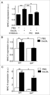
SYK regulates macrophage MHC-II expression in response to OxLDL. (A) BMDM isolated from C57BL/6 mice were pretreated with 40 μM SYK inhibitor (PIC, piceatannol) or 1 mM autophagy inhibitor (3MA) for 2 h and then incubated with PBS or 25 μg/ml OxLDL for additional 18 h. MHC-II expression was measured by FACS. (B) BMDM isolated from WT and syk−/− mice were incubated with PBS or 25 μg/ml OxLDL for 18 h and surface MHC-II expression was measured by FACS. (C) WT and syk−/− mice were intraperitoneally injected with 0.2 ml of PBS or 100 μg/ml OxLDL. After 24 h, peritoneal cells were isolated and MHC-II expression on F4/80-positive macrophages was measured by FACS. Mean± SE ; n = 3 −4. *, P < 0.05; **, P < 0.005.
Figure 7.
Figure 7.
Macrophage SYK expression is required for antigen presentation to CD4+ T cells. WT- or SYK-deficient BMDM were pretreated with 25 μg/ml OxLDL plus 25 μg/ml MDA-LDL for 18 h and then cocultured with CD4+ T cells isolated from the spleens of WT mice immunized with MDA-LDL for additional 72 h. (A and B) At 72 h, T cell proliferation was measured by FACS. (C) At 48 h, IL2 levels in coculture media were measured by ELISA. Mean± SE ; n = 3 −4. *, P < 0.05; **, P < 0.005.
Figure 8.
Figure 8.
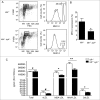
Macrophage SYK expression regulates OSE-specific IgG levels in hypercholesterolemic mice. ldlr−/− and ldlr−/− syk−/− mice were fed a HFD for 12 wk. (A) Peritoneal cells were isolated and MHC-II expression on F4/80-positive macrophages was measured by FACS. (B) Quantification of the results presented in (A). Mean±SE ; ldlr−/−: n=5, ldlr−/− syk−/−: n=8 ; *, P < 0.05. (C) Plasma was isolated from HFD-fed ldlr−/− and ldlr−/− syk−/− mice. Levels of total IgG2c and the IgG2c against native and oxidatively modified LDL were measured by ELISA. Mean± SE ; ldlr−/−: n = 13, ldlr−/− syk−/−: n = 18 . #, P > 0.05; *, P < 0.05; **, P < 0.005. Note that one cannot compare absolute values for total and antigen-specific titers in these assays as different conditions were used.

References

    1. Hansson GK, Libby P. The immune response in atherosclerosis: a double-edged sword. Nat Rev Immunol 2006; 6:508-19; PMID:16778830; http://dx.doi.org/10.1038/nri1882 - DOI - PubMed
    1. Lichtman AH, Binder CJ, Tsimikas S, Witztum JL. Adaptive immunity in atherogenesis: new insights and therapeutic approaches. J Clin Invest 2013; 123:27-36; PMID:23281407; http://dx.doi.org/10.1172/JCI63108 - DOI - PMC - PubMed
    1. Palinski W, Horkko S, Miller E, Steinbrecher UP, Powell HC, Curtiss LK, Witztum JL. Cloning of monoclonal autoantibodies to epitopes of oxidized lipoproteins from apolipoprotein E-deficient mice. demonstration of epitopes of oxidized low density lipoprotein in human plasma. J Clin Invest 1996; 98:800-14; PMID:8698873; http://dx.doi.org/10.1172/JCI118853 - DOI - PMC - PubMed
    1. Binder CJ, Hartvigsen K, Chang MK, Miller M, Broide D, Palinski W, Curtiss LK, Corr M, Witztum JL. IL-5 links adaptive and natural immunity specific for epitopes of oxidized LDL and protects from atherosclerosis. J Clin Invest 2004; 114:427-37; PMID:15286809; http://dx.doi.org/10.1172/JCI200420479 - DOI - PMC - PubMed
    1. Gonen A, Hansen LF, Turner WW, Montano EN, Que X, Rafia A, Chou MY, Wiesner P, Tsiantoulas D, Corr M, et al.. Atheroprotective immunization with malondialdehyde-modified LDL is hapten specific and dependent on advanced MDA adducts: implications for development of an atheroprotective vaccine. J Lipid Res 2014; 55:2137-55; PMID:25143462; http://dx.doi.org/10.1194/jlr.M053256 - DOI - PMC - PubMed

Publication types

MeSH terms