

Free-breathing liver perfusion imaging using 3-dimensional through-time spiral generalized autocalibrating partially parallel acquisition acceleration
- PMID: 25946703
- PMCID: PMC4423561
- DOI: 10.1097/RLI.0000000000000135
Free-breathing liver perfusion imaging using 3-dimensional through-time spiral generalized autocalibrating partially parallel acquisition acceleration
Abstract
Objectives: The goal of this study was to develop free-breathing high-spatiotemporal resolution dynamic contrast-enhanced liver magnetic resonance imaging using non-Cartesian parallel imaging acceleration, and quantitative liver perfusion mapping.
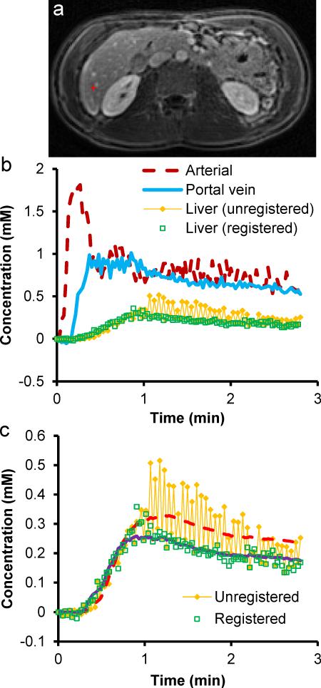
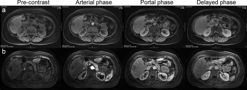
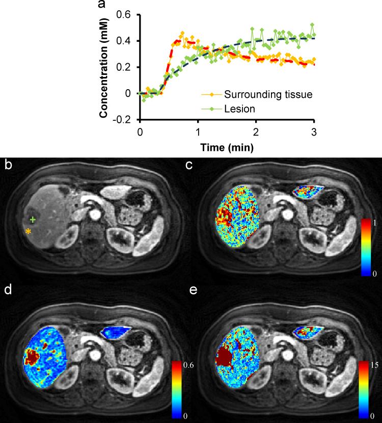
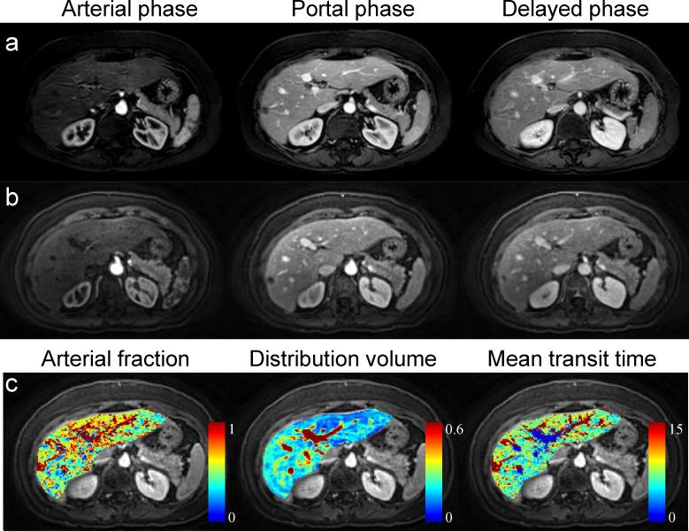
Materials and methods: This study was approved by the local institutional review board and written informed consent was obtained from all participants. Ten healthy subjects and 5 patients were scanned on a Siemens 3-T Skyra scanner. A stack-of-spirals trajectory was undersampled in-plane with a reduction factor of 6 and reconstructed using 3-dimensional (3D) through-time non-Cartesian generalized autocalibrating partially parallel acquisition. High-resolution 3D images were acquired with a true temporal resolution of 1.6 to 1.9 seconds while the subjects were breathing freely. A dual-input single-compartment model was used to retrieve liver perfusion parameters from dynamic contrast-enhanced magnetic resonance imaging data, which were coregistered using an algorithm designed to reduce the effects of dynamic contrast changes on registration. Image quality evaluation was performed on spiral images and conventional images from 5 healthy subjects.
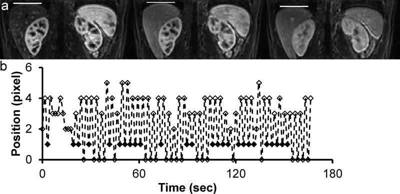
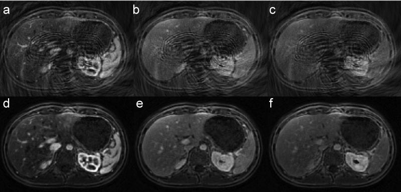
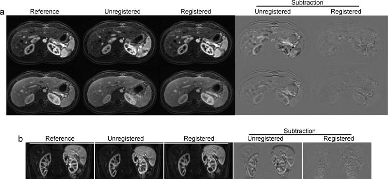
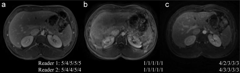
Results: Images with a spatial resolution of 1.9 × 1.9 × 3 mm3 were obtained with whole-liver coverage. With an imaging speed of better than 2 s/vol, free-breathing scans were achieved and dynamic changes in enhancement were captured. The overall image quality of free-breathing spiral images was slightly lower than that of conventional long breath-hold Cartesian images, but it provided clinically acceptable or better image quality. The free-breathing 3D images were registered with almost no residual motion in liver tissue. After the registration, quantitative whole-liver 3D perfusion maps were obtained and the perfusion parameters are all in good agreement with the literature.
Conclusions: This high-spatiotemporal resolution free-breathing 3D liver imaging technique allows voxelwise quantification of liver perfusion.
Figures
References
-
- Hanley J, Debois MM, Mah D, et al. Deep inspiration breath-hold technique for lung tumors: the potential value of target immobilization and reduced lung density in dose escalation. Int. J. Radiat. Oncol. Biol. Phys. 1999;45(3):603–611. - PubMed
-
- Riffel P, Attenberger UI, Kannengiesser S, et al. Highly accelerated T1-weighted abdominal imaging using 2-dimensional controlled aliasing in parallel imaging results in higher acceleration: a comparison with generalized autocalibrating partially parallel acquisitions parallel imaging. Invest. Radiol. 2013;48(7):554–561. - PubMed
-
- Michaely HJ, Morelli JN, Budjan J, et al. CAIPIRINHA-Dixon-TWIST (CDT)-volume-interpolated breath-hold examination (VIBE): a new technique for fast time-resolved dynamic 3-dimensional imaging of the abdomen with high spatial resolution. Invest. Radiol. 2013;48(8):590–597. - PubMed
-
- Park YS, Lee CH, Kim IS, et al. Usefulness of controlled aliasing in parallel imaging results in higher acceleration in gadoxetic acid-enhanced liver magnetic resonance imaging to clarify the hepatic arterial phase. Invest. Radiol. 2014;49(3):183–188. - PubMed
Publication types
MeSH terms
Substances
Grants and funding
LinkOut - more resources
Full Text Sources
Other Literature Sources
Medical
