

VALIDATION OF THERAPEUTIC RESPONSE ASSESSMENT BY BONE SCINTIGRAPHY IN PATIENTS WITH BONE-ONLY METASTATIC BREAST CANCERS DURING ZOLEDRONIC ACID TREATMENT: COMPARISON WITH COMPUTED TOMOGRAPHY ASSESSMENT
- PMID: 25946906
- PMCID: PMC5131596
- DOI: 10.5387/fms.2013-15
VALIDATION OF THERAPEUTIC RESPONSE ASSESSMENT BY BONE SCINTIGRAPHY IN PATIENTS WITH BONE-ONLY METASTATIC BREAST CANCERS DURING ZOLEDRONIC ACID TREATMENT: COMPARISON WITH COMPUTED TOMOGRAPHY ASSESSMENT
Abstract
Purpose: To validate the use of bone scintigraphy (BS) versus computed tomography (CT) for therapeutic monitoring in patients during treatment with zoledronic acid.
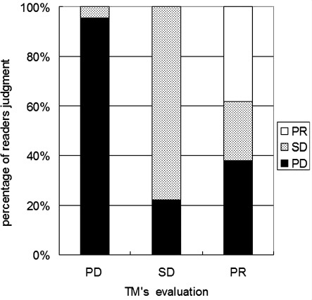
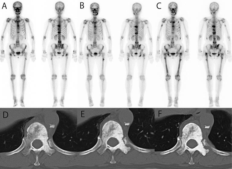
Materials and methods: Eleven patients with bone-only metastatic disease and being treated with zoledronic acid were included. The effects of therapies including chemotherapy and hormone therapy were evaluated in 25 separate examinations in total as follows: complete response (CR), when no bone metastasis was visible; partial response (PR), when a decrease in the lesion area was detected; stable disease (SD), when no or slight change was observed; and progressive disease (PD), when new or enlarged lesion areas were observed.
Results: The accuracies of examination by Readers 1, 2, and 3 respectively were 76%, 80% and 76% for BS, 52%, 48%, and 40% for CT, and 64%, 52% and 60% for BS and CT combined with Readers 2 and 3 observing significant differences between CT and BS results. The rates of interobserver agreement between Readers 1 and 2, between Readers 1 and 3, and between Reader 2 and 3 respectively, were 84%, 80% and 88% (κ = 0.648, 0.561 and 0.766) for BS, 52%, 56%, and 60% (κ = 0.180, 0.278 and 0.282) for CT, and 52%, 60%, and 56% (κ = 0.215, 0.282 and 0.232) for CT and BS combined.
Conclusion: BS is effective for assessing the response of bone metastasis to therapy in patients during zoledronic acid treatment.
Figures
Similar articles
-
Is retention of zoledronic acid onto bone different in multiple myeloma and breast cancer patients with bone metastasis?J Bone Miner Res. 2013 Aug;28(8):1738-50. doi: 10.1002/jbmr.1897. J Bone Miner Res. 2013. PMID: 23427025 Clinical Trial.
-
Phase II trial evaluating the palliative benefit of second-line zoledronic acid in breast cancer patients with either a skeletal-related event or progressive bone metastases despite first-line bisphosphonate therapy.J Clin Oncol. 2006 Oct 20;24(30):4895-900. doi: 10.1200/JCO.2006.05.9212. Epub 2006 Sep 25. J Clin Oncol. 2006. PMID: 17001071 Clinical Trial.
-
Comparison of (18)F-FDG-PET/CT with (99m)Tc-MDP bone scintigraphy for the detection of bone metastases in cancer patients.Nucl Med Commun. 2010 Jun;31(6):597-603. doi: 10.1097/MNM.0b013e328338e909. Nucl Med Commun. 2010. PMID: 20224457
-
Zoledronic acid in the treatment of metastatic breast cancer.Anticancer Drugs. 2014 Jan;25(1):1-7. doi: 10.1097/CAD.0000000000000020. Anticancer Drugs. 2014. PMID: 24100278 Review.
-
Clinical efficacy and safety of zoledronic acid in prostate and breast cancer.Expert Rev Anticancer Ther. 2009 Sep;9(9):1211-8. doi: 10.1586/era.09.95. Expert Rev Anticancer Ther. 2009. PMID: 19761424 Review.
References
-
- Maffioli L, Florimonte L, Pagani L, Butti I, Roca I. Current role of bone scan with phosphonates in the follow-up of breast cancer. Eur J Nucl Med Mol Imaging, 31: S143-148, 2004. - PubMed
-
- Dhillon S, Lyseng-Williamson KA. Zoledronic acid: a review of its use in the management of bone metastases of malignancy. Drugs, 68: 507-534, 2008. - PubMed
-
- Green JR. Antitumor effects of bisphosphonates. Cancer, 97 (Suppl): 840- 847, 2003. - PubMed
-
- Mundy GR, Yoneda T, Hiraga T. Preclinical studies with zoledronic acid and other bisphosphonates: impact on the bone microenvironment. Semin Oncol, 28 (Suppl): 35- 44, 2001. - PubMed
Publication types
MeSH terms
Substances
LinkOut - more resources
Full Text Sources
Other Literature Sources
Medical
Research Materials