. 2016 Apr;65(4):635-46.
doi: 10.1136/gutjnl-2014-308257. Epub 2015 May 6.

Increased expression of Solute carrier family 12 member 5 via gene amplification contributes to tumour progression and metastasis and associates with poor survival in colorectal cancer

Affiliations

Increased expression of Solute carrier family 12 member 5 via gene amplification contributes to tumour progression and metastasis and associates with poor survival in colorectal cancer

Lixia Xu et al. Gut. 2016 Apr.

Abstract

Objective: Using whole genome sequencing, we identified gene amplification of solute carrier family 12 member 5 (SLC12A5) located at 20q13.12 in colorectal cancer (CRC). We analysed its amplification, overexpression, biological effects and prognostic significance in CRC.

Design: SLC12A5 amplification status was evaluated by fluorescence in situ hybridisation (FISH). The effects of SLC12A5 re-expression or knockdown were determined in proliferation, apoptosis, invasion and metastasis assays. SLC12A5 target genes and related pathways were identified by reporter activity and cDNA microarray analyses. Clinical impact of SLC12A5 overexpression was assessed in 195 patients with CRC.

Results: Amplification of SLC12A5 was verified in 78 out of 191 (40.8%) patients with primary CRC by FISH, which was positively correlated with its protein overexpression (p<0.001). Biofunctional investigation of SLC12A5 revealed that SLC12A5 significantly increased cell proliferation, G1-S cell cycle transition, invasion/migration abilities, but suppressed apoptosis in vitro and promoted xenograft tumour growth as well as lung metastasis in vivo. The antiapoptosis effect by SLC12A5 was mediated through inhibiting apoptosis-inducing factor and endonuclease G-dependent apoptotic signalling pathway; and the pro-metastasis role was by regulating key elements of the matrix architecture, including matrix metallopeptidase and fibronectin. After a median follow-up of 50.16 months, multivariate analysis revealed that patients with SLC12A5 protein overexpression had a significant decrease in overall survival. Kaplan-Meier survival curves showed that SLC12A5 overexpression was significantly associated with shortened survival in patients with CRC.

Conclusions: SLC12A5 plays a pivotal oncogenic role in colorectal carcinogenesis; its overexpression is an independent prognostic factor of patients with CRC.

Keywords: COLORECTAL CANCER; GENE EXPRESSION; MOLECULAR ONCOLOGY; TUMOUR MARKERS.


Figures

Figure 1
Figure 1
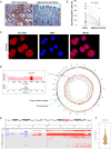
SLC12A5 is amplified in colorectal cancer (CRC). (A) Representative images of SLC12A5 protein expression in CRC tumour tissues and their adjacent tissues by immunohistochemistry. (B) The protein expression level of SLC12A5 was significantly higher in primary CRC tumour as compared with their adjacent tissue (p<0.05). (C) SLC12A5 was expressed in both nuclear and cytoplasm in colon cancer cell line HCT116 by immunofluorescence and confocal microscopy. (D) The right panel shows the genomic profiles of CRC by whole genome sequencing, where the copy number alterations were indicated with red bars. The left panel shows the visualisation of copy ratio particularly for the whole chromosome 20. 20q13.12 is highlighted with red colour, which harbours the amplified gene SLC12A5. (E) Significant genomic alterations of 20q13.12 across 12 human cancer types in The Cancer Genome Atlas (TCGA) cohort. Red colour indicates copy number gain, while blue colour refers to copy number loss. BLCA, bladder urothelial carcinoma; BRCA, breast invasive carcinoma; KIRC, kidney renal clear cell carcinoma; COAD, colon adenocarcinoma; DAPI, 4’,6-diamidino-2-phenylindole; GBM, glioblastoma multiforme; HNSC, head and neck squamous cell carcinoma; LAML, acute myeloid leukaemia; LUAD, lung adenocarcinoma; LUSC, lung squamous cell carcinoma; READ, rectum adenocarcinoma; OV, ovarian serous cystadenocarcinoma; UCEC, uterine corpus endometrioid carcinoma. (F) Copy number changes of SLC12A5 in 575 colorectal cancers from TCGA.
Figure 2
Figure 2
Amplification and overexpression of SLC12A5 in colorectal cancers (CRCs). (A) Fluorescence in situ hybridisation (FISH) was performed in normal metaphase lymphocytes and colon cancer cell line Caco-2. The red signal indicates the location of SLC12A5 and the green signal refers to centromere of chromosome 20. This result implied that the probe of SLC12A5 was correctly located on chromosome 20. Amplification of SLC12A5 was observed in Caco-2. (B) Representative image of SLC12A5 gene amplification detected by FISH in primary CRC tissues, but not detected in their adjacent tissues. (C) Representative staining of high and low expression levels of SLC12A5 in CRC tissue microarray slides by immunohistochemistry. (D) The correlation between copy number changes of SLC12A5 and protein overexpression (R2=0.506, p<0.001).
Figure 3
Figure 3
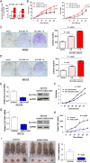
SLC12A5 promotes colorectal cancer cell growth. (A) Expression levels of SLC12A5 were increased after transfection with different doses of SLC12A5 plasmids (0.8 and 1.6 µg) in colon cancer cell lines SW480 and SW1116. (B) Ectopic expression of SLC12A5 significantly enhanced cell viability in a dose-dependent manner in both cell lines. (**p<0.01, ***p<0.001) (C) Empty vector and different doses of SLC12A5 plasmids (0.8 and 1.6 µg) were used to transfect cells in 24-well plates. Compared with empty vector-transfected cells, colony numbers significantly increased with transfection of 0.8 and 1.6 µg SLC12A5 plasmids in SW480 in a dose-dependent manner. (D) The number of colonies increased in a dose-dependent manner when transfected with SLC12A5 plasmids in SW1116. (E) Knockdown efficiency of SLC12A5 in HCT116 cells by transient transfection of siSLC12A5 was examined by real-time PCR (left panel) and western blot (right panel), respectively. (F) Knockdown of SLC12A5 significantly inhibited cell viability in HCT116. (G) Knockdown efficiency of SLC12A5 in HCT116 cells by stable transfection of shSLC12A5 was demonstrated by real-time PCR (left panel) and western blot (right panel). (H) The tumour growth curve of HCT116 stably transduced with shSLC12A5 in nude mice was significantly dampened compared with HCT116 transduced with Control. (I) A representative picture of tumour formation in nude mice subcutaneously inoculated with shSLC12A5- or Control-HCT116 (left panel). Histogram represents mean of the tumour weight from the shSLC12A5 and Control groups (right panel).
Figure 4
Figure 4
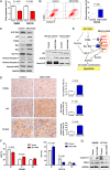
SLC12A5 reduces apoptosis in colorectal cancer cell lines. (A) Overexpression of SLC12A5 significantly inhibited early apoptosis in SW480 and SW1116 (B) Knockdown of SLC12A5 significantly increased the proportion of apoptotic cells in HCT116 cells by flow cytometry analyses following Annexin V and 7-amino-actinomycin (7-AAD) staining (p<0.01). (C) Protein expression of key apoptosis-related genes was evaluated by western blot in HCT116 cells transfected with siSLC12A5 or Control. After silencing SLC12A5 expression, no changes of cleaved Caspase 3, 7 nor cleaved poly(ADP-ribose) polymerase (PARP) were observed. SLC12A5 silencing increased the protein expression of p53, p53 upregulated modulator of apoptosis (PUMA), Bax and Bak and downregulated the inhibitor of apoptosis protein Survivin. Knockdown of SLC12A5 enhanced the nuclear protein levels of apoptosis-inducing factor (AIF) and endonuclease G (EndoG). (D) TUNEL-stained sections of mouse xenograft tumours displayed apoptotic cells; representative images of AIF and EndoG immunohistochemistry-stained sections of mouse xenograft tumours. (E) Schematic diagram for the proposed mechanisms of SLC12A5 exerting antiapoptotic function via caspase-independent signalling pathway. (F) The number of cell distribution was determined by flow cytometry. Values are mean±SD. (G) Protein expression of cyclin D1 and cyclin-dependent kinase (CDK4) was determined by western blot.
Figure 5
Figure 5
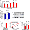
SLC12A5 suppressed p53 and p21 luciferase reporter activity. (A) To screen for SLC12A5 target signalling pathways, a serial of promoter-luciferase assays (p53-luc, p21-luc, activator protein 1 (AP1)-luc, nuclear factor κB (NF κB)-luc, and Wnt/β-catenin) were performed in SLC12A5 stably transfected p53 wildtype (WT) HCT116 cells compared with vector control cells. Luciferase activities were determined by dual luciferase assay system at 48 h post-transfection. Ectopic expression of SLC12A5 suppressed p53 and p21 luciferase reporter activity in p53 WT HCT116. (B) Knockdown of SLC12A5 increased p53 and p21 luciferase reporter activity in p53 WT HCT116. (C) The western blot results confirmed the findings of luciferase reporter activity assays in p53 WT HCT116. (D) Ectopic expression of SLC12A5 was determined in p53 knockout (KO) HCT116 cells by real-time PCR. (E) Ectopic expression of SLC12A5 significantly promoted cell growth in p53 KO HCT116 cells. (F) SLC12A5 in HCT116 cells suppressed p21-luciferase reporter activity in both p53 KO HCT116 and SW116 cells.
Figure 6
Figure 6
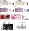
SLC12A5 promotes tumour metastasis in vitro and in vivo. (A) Ectopic expression of SLC12A5 promoted cell invasion and migration in SW1116 cells. (B) Knockdown of SLC12A5 suppressed cell invasion and migration in HCT116 cells. (C) Representative images of lungs with or without metastasis (left panel). H&E staining of lung tissues from nude mice injected with Control-HCT116 or shSLC12A5-HCT116 cells (200x magnification, middle panel). Incidence of lung metastasis (right panel). (D) Validation of the key metastasis-related genes identified by cDNA expression array by RT-PCR. (E) Western blot further confirmed the deregulation of metastasis-related genes. GAPDH, glyceraldehyde 3-phosphate dehydrogenase; HGF, hepatocyte growth factor; MMP, matrix metallopeptidase.
Figure 7
Figure 7
Clinical significance of SLC12A5 in colorectal cancers (CRCs). Kaplan–Meier survival analysis according to SLC12A5 expression in195 patients with CRC. Patients with CRC with SLC12A5 overexpression had poorer survival than others. The difference is statistically significant based on the log-rank test (p=0.009).

Similar articles

Cited by

References

    1. Jemal A, Bray F, Center MM. , et al. Global cancer statistics. CA Cancer J Clin 2011;61:69–90. 10.3322/caac.20107 - DOI - PubMed
    1. Sung JJ, Lau JY, Young GP. , et al. Asia Pacific consensus recommendations for colorectal cancer screening. Gut 2008;57:1166–76. 10.1136/gut.2007.146316 - DOI - PubMed
    1. Lubbe WJ, Zuzga DS, Zhou Z. , et al. Guanylyl cyclase C prevents colon cancer metastasis by regulating tumor epithelial cell matrix metalloproteinase-9. Cancer Res 2009;69:3529–36. 10.1158/0008-5472.CAN-09-0067 - DOI - PMC - PubMed
    1. Rokavec M, Oner MG, Li H. , et al. IL-6R/STAT3/miR-34a feedback loop promotes EMT-mediated colorectal cancer invasion and metastasis. J Clin Invest 2014;124:1853–67. 10.1172/JCI73531 - DOI - PMC - PubMed
    1. Fearon ER. Molecular genetics of colorectal cancer. Annu Rev Pathol 2011;6:479–507. 10.1146/annurev-pathol-011110-130235 - DOI - PubMed

Publication types

MeSH terms

Substances