Comparative Study
. 2015 May 19;112(20):E2695-704.
doi: 10.1073/pnas.1410767112. Epub 2015 May 6.

Connectivity reveals relationship of brain areas for reward-guided learning and decision making in human and monkey frontal cortex

Affiliations
Comparative Study

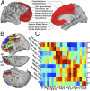
Connectivity reveals relationship of brain areas for reward-guided learning and decision making in human and monkey frontal cortex

Franz-Xaver Neubert et al. Proc Natl Acad Sci U S A. .

Abstract

Reward-guided decision-making depends on a network of brain regions. Among these are the orbitofrontal and the anterior cingulate cortex. However, it is difficult to ascertain if these areas constitute anatomical and functional unities, and how these areas correspond between monkeys and humans. To address these questions we looked at connectivity profiles of these areas using resting-state functional MRI in 38 humans and 25 macaque monkeys. We sought brain regions in the macaque that resembled 10 human areas identified with decision making and brain regions in the human that resembled six macaque areas identified with decision making. We also used diffusion-weighted MRI to delineate key human orbital and medial frontal brain regions. We identified 21 different regions, many of which could be linked to particular aspects of reward-guided learning, valuation, and decision making, and in many cases we identified areas in the macaque with similar coupling profiles.

Keywords: anterior cingulate cortex; comparative anatomy; decision making; orbitofrontal cortex; resting state functional connectivity.


Conflict of interest statement

The authors declare no conflict of interest.

Figures

Fig. 1.
Fig. 1.
(A) Overall approach of the study. fMRI analyses in 38 humans and 25 macaques were used to establish the whole-brain functional connectivity of regions in medial and orbital frontal cortex identified with reward-guided learning and decision making in the two species. The example shows the macaque brain regions that have a similar coupling profile to a human vmPFC region identified in a decision-making study (27). Reproduced from ref. , with permission from Macmillan Publishers Ltd, Nature Neuroscience. (B) Each region’s functional connectivity with 23 key regions was then determined and (C) summarized as a functional connectivity fingerprint. (D) Once the functional connectivity fingerprint of a human brain area was established it was compared with the functional connectivity fingerprints of 380 ROIs in macaque orbital and medial frontal cortex (one example is shown here) by calculating the summed absolute difference [the “Manhattan” or “city-block” distance (–19) of the coupling scores]. (E) Examples of the functional connectivity fingerprints for a human (blue) and a monkey (red) brain area. Most monkey ROIs matched human areas relatively poorly and extremely good and extremely bad matches were relatively rare. We used two SDs below the mean of this distribution of summed absolute differences as a cut-off to look for “significantly” good human to monkey matches. (F) A heat map summarizing the degree of correspondence between the functional connectivity patterns of each voxel in the macaque and the human brain region shown in A. Warm red areas indicate macaque voxels that correspond most strongly. (G) Complementary parts of the investigation started with the functional connectivity fingerprints of both human (Upper) and macaque (Lower) brain areas involved in reward-guided learning and decision making and then compared them with the functional connectivity fingerprints of areas in the other species. (Top Left) Reproduced from ref. , with permission from Macmillan Publishers Ltd, Nature Neuroscience. (Bottom Left) Reproduced from ref. , with permission from Macmillan Publishers Ltd, Nature Neuroscience.
Fig. 2.
Fig. 2.
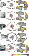
Human medial frontal regions (Left) linked with (A) reward-guided decision making (21), (B) more abstract reward-guided decision making (28), (C) cost-benefit valuation (27), (D) imagining the reward outcomes of others (40), and (E) self-valuation and depression (42), could all be linked to macaque brain regions (Right) via similarities in their coupling patterns (Center: blue, macaque; red, human). However, a human brain area (D) associated with reward outcome imagination did not correspond in a simple way with any area in the macaque. (A) Reproduced from ref. , with permission from Elsevier. (B) Reproduced from ref. , with permission from Macmillan Publishers Ltd, Nature Neuroscience. (C) Reproduced from ref. , with permission from Macmillan Publishers Ltd, Nature Neuroscience. (D) Modified from ref. . (E) Modified from ref. .
Fig. 3.
Fig. 3.
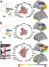
Macaque medial frontal regions (Left) associated with (A) positive reward expectations (5), (B) negative outcome expectations (5), and (C) cost-benefit decision making (11) could all be linked to human brain regions (Right) via similarities in their coupling patterns (Center). (A and B) Reproduced from ref. . (C) Modified from ref. , with permission from Macmillan Publishers Ltd, Nature Neuroscience.
Fig. 4.
Fig. 4.
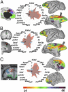
Human ACC regions (Left) linked with (A) cognitive control (13) and (B) reward-guided action selection and behavioral updating (49) could be linked to macaque brain regions (Right) via similarities in their coupling patterns (Center). (C) A macaque ACC region (Left) linked with reward-guided behavioral updating (51) could be linked to a similar human ACC region to that shown in B. (A) Modified from ref. , with permission from Elsevier. (B) Reproduced from ref. , with permission from Macmillan Publishers Ltd, Nature Neuroscience. (C) Reproduced from ref. , with permission from Macmillan Publishers Ltd, Nature Neuroscience.
Fig. 5.
Fig. 5.
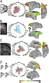
Human OFC regions (Left) linked with (A) stimulus-reward association (55) and could be linked to a macaque lOFC (Right) via similarities in the regions’ coupling patterns (Center). (B) A human brain concerned with decision-making under ambiguity (61) did not correspond strongly to any macaque brain region. (C and D) Macaque regions (Left) linked with (C) context-independent and outcome identity-dependent value signals (53) and (D) decision outcome monitoring (63) could be linked to human brain regions (Right) via similarities in coupling patterns (Center). (A) Reproduced from ref. . (B) Modified from ref. . (C) Reproduced from ref. , with permission from Macmillan Publishers Ltd, Nature Neuroscience. (D) Modified from ref. , with permission from Macmillan Publishers Ltd, Nature Neuroscience.
Fig. 6.
Fig. 6.
(A) The human medial and orbital region investigated and (B) the subregions that could be identified on the basis of differences in DW-MRI–estimated connectivity. (C) Correspondences between the fMRI-based functional coupling maps associated with decision-making areas (Figs. 1–5) and orbital and medial subregions identified via DW-MRI parcellation (B). Warm red colors indicate similarities in the functional coupling maps and asterisks indicate areas with highest spatial correlation (see SI Appendix, Section 5 for more information).

References

    1. Wallis JD. Cross-species studies of orbitofrontal cortex and value-based decision-making. Nat Neurosci. 2012;15(1):13–19. - PMC - PubMed
    1. Rangel A, Hare T. Neural computations associated with goal-directed choice. Curr Opin Neurobiol. 2010;20(2):262–270. - PubMed
    1. O’Doherty JP. Contributions of the ventromedial prefrontal cortex to goal-directed action selection. Ann N Y Acad Sci. 2011;1239:118–129. - PubMed
    1. Bouret S, Richmond BJ. Ventromedial and orbital prefrontal neurons differentially encode internally and externally driven motivational values in monkeys. J Neurosci. 2010;30(25):8591–8601. - PMC - PubMed
    1. Monosov IE, Hikosaka O. Regionally distinct processing of rewards and punishments by the primate ventromedial prefrontal cortex. J Neurosci. 2012;32(30):10318–10330. - PMC - PubMed

Publication types