. 2015 Jun 19;290(25):15825-15834.
doi: 10.1074/jbc.M115.648428. Epub 2015 May 6.

The Tyrosine Kinase c-Src Specifically Binds to the Active Integrin αIIbβ3 to Initiate Outside-in Signaling in Platelets

Affiliations

The Tyrosine Kinase c-Src Specifically Binds to the Active Integrin αIIbβ3 to Initiate Outside-in Signaling in Platelets

Yibing Wu et al. J Biol Chem. .

Abstract

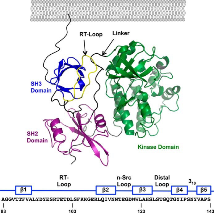
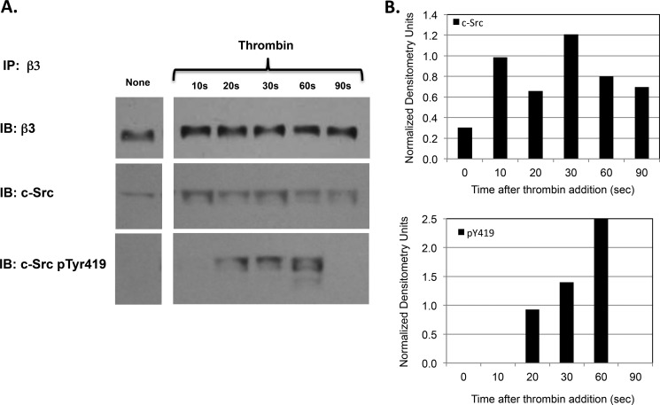
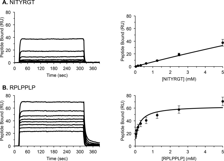
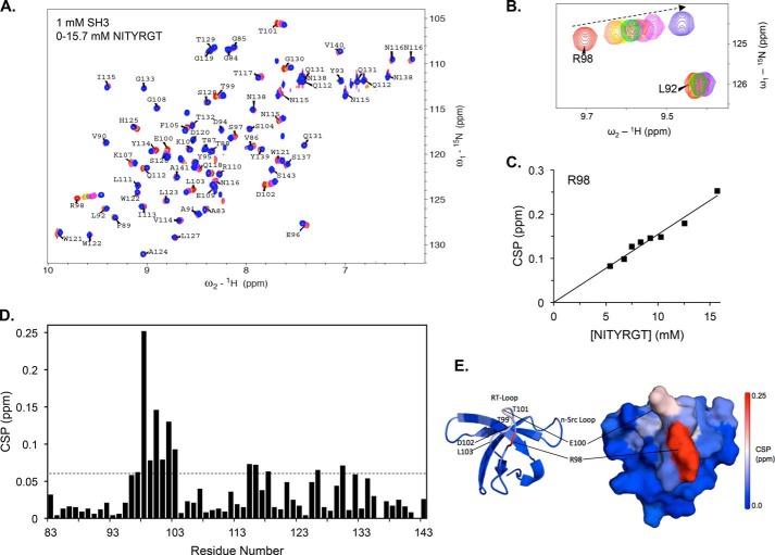
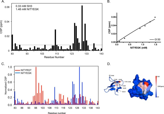
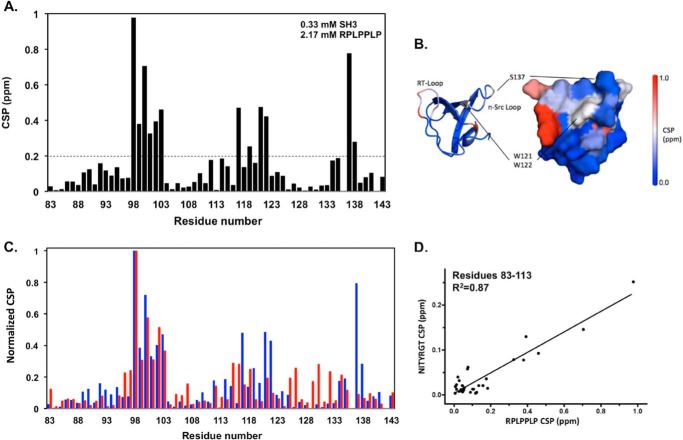
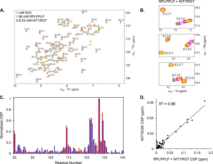
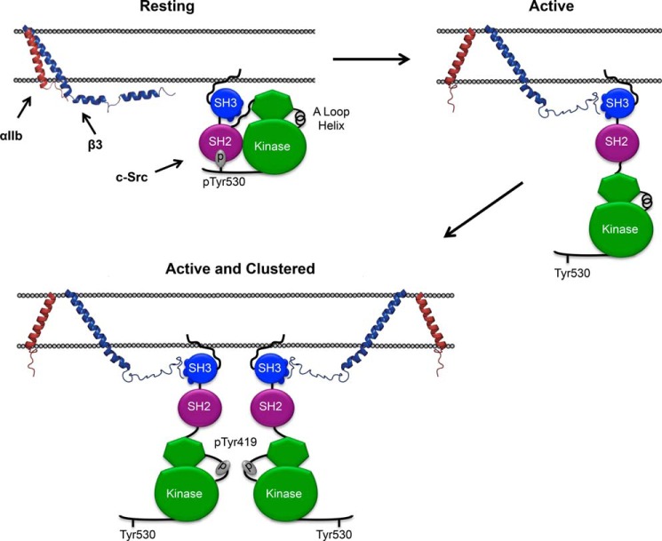
It is currently believed that inactive tyrosine kinase c-Src in platelets binds to the cytoplasmic tail of the β3 integrin subunit via its SH3 domain. Although a recent NMR study supports this contention, it is likely that such binding would be precluded in inactive c-Src because an auto-inhibitory linker physically occludes the β3 tail binding site. Accordingly, we have re-examined c-Src binding to β3 by immunoprecipitation as well as NMR spectroscopy. In unstimulated platelets, we detected little to no interaction between c-Src and β3. Following platelet activation, however, c-Src was co-immunoprecipitated with β3 in a time-dependent manner and underwent progressive activation as well. We then measured chemical shift perturbations in the (15)N-labeled SH3 domain induced by the C-terminal β3 tail peptide NITYRGT and found that the peptide interacted with the SH3 domain RT-loop and surrounding residues. A control peptide whose last three residues where replaced with those of the β1 cytoplasmic tail induced only small chemical shift perturbations on the opposite face of the SH3 domain. Next, to mimic inactive c-Src, we found that the canonical polyproline peptide RPLPPLP prevented binding of the β3 peptide to the RT- loop. Under these conditions, the β3 peptide induced chemical shift perturbations similar to the negative control. We conclude that the primary interaction of c-Src with the β3 tail occurs in its activated state and at a site that overlaps with PPII binding site in its SH3 domain. Interactions of inactive c-Src with β3 are weak and insensitive to β3 tail mutations.

Keywords: Src; Src homology 3 domain (SH3 domain); integrin; platelet; signal transduction.


Figures

FIGURE 1.
FIGURE 1.
A, domain structure of inactive c-Src (PDB 2SRC). From N- to C terminus: SH3 domain (blue); SH2 domain (purple); kinase domain (green). The linker between the SH2 and kinase domains is colored black, and the RT-loop in the SH3 domain is colored yellow. The amino acid sequence of the human c-Src SH3 domain is numbered according to UniProtKB/Swiss-Prot entry P12931 and indicates the location of its five β strands and the interconnecting loops.
FIGURE 2.
FIGURE 2.
A, time course of c-Src binding to the integrin β3 subunit and its subsequent activation after platelet stimulation by thrombin. Following the stimulation of washed human platelets by 1 unit/ml thrombin for the indicated times, platelets were lysed with 1% Nonidet P-40, β3 was immunoprecipitated (IP) from the lysates using a β3-specific monoclonal antibody, and the immunoprecipitated β3 was serially immunoblotted (IB) for β3, c-Src, and phosphorylated c-Src residue 419 (pTyr419). B, densitometry using NIH ImageJ software of the immunoblot shown in Fig. 2A demonstrating the time course of c-Src binding to the integrin β3 subunit and its subsequent activation after platelet stimulation by thrombin. The densitometry for β3-bound c-Src and for β3-bound c-Src containing phosphorylated tyrosine residue 419 (pY419) at each time point was normalized using the densitometry for the corresponding β3 band.
FIGURE 3.
FIGURE 3.
Representative SPR measurements of steady state NITYRGT (A) and RPLPPLP (B) binding to the c-Src SH3 domain. Average dissociation constants (Kd), calculated using BIAevaluation software, from 9–12 steady state measurements, were 259 ± 52 μm and 320 ± 120 mm for RPLPPLP and NITYRGT, respectively.
FIGURE 4.
FIGURE 4.
The C-terminal β3 peptide NITYRGT binds to the RT-Loop of the c-Src SH3 domain. A, superimposition of HSQC spectra of 1 mm 15N-labeled c-Src SH3 domain in the presence of 0, 5.4, 6.7, 7.5, 8.3, 9.2, 10.3, 12.6, and 15.7 mm NITYRGT. B, subspectra for c-Src SH3 domain residues Leu-92 and Arg-98 at NITYRGT concentrations of 0, 5.4, 6.7, 8.3, 9.2, 10.3, 12.6, and 15.7 mm. C, plot of CSP in ppm for residue Arg-98 as a function of NITYRGT concentration. D, CSP induced in each residue of the c-Src SH3 domain by 15.7 mm NITYRGT. The dotted line represents 2 standard deviations for CSP induced in the c-Src SH3 HSQC, determined by iteratively fitting the CSP to normal distributions as described by Schumann et al. (26). The calculated standard deviation of NITYRGT-induced CSP was 0.027 ppm. E, CSP induced by NITYRGT in the c-Src SH3 domain are color-coded by magnitude and mapped onto an NMR structure of the SH3 domain (PDB 1NLP).
FIGURE 5.
FIGURE 5.
Interaction of the mutant β3/β1 peptide NITYEGK with the c-Src SH3 domain. A, CSP induced in each residue of the c-Src SH3 domain by NITYEGK. The dotted line represents 2 standard deviations for NITYEGK-induced CSP. The calculated standard deviation of NITYEGK-induced CSP was 0.004 ppm. B, plot of the magnitude of CSP in ppm for residue Gly-130 as a function of NITYEGK concentration. C, superimposition of normalized CSP induced by NITYEGK (blue) and NITYRGT (red, taken from Fig. 4). NITYEGK CSP were normalized by dividing the maximal CSP for each SH3 domain residue in NITYEGK spectra by the maximal CSP for Gly-130. NITYRGT CSP were normalized by dividing the maximal CSP for each residue in the NITYRGT spectra by the maximal CSP for Arg-98. D, CSP induced in the c-Src SH3 domain by NITYEGK are color-coded by magnitude and mapped onto an NMR structure of the SH3 domain (PDB 1NLP).
FIGURE 6.
FIGURE 6.
NITYRGT and the PPII core peptide RPLPPLP bind to the same region of the c-Src SH3 domain. A, CSP caused by the peptide RPLPPLP for each residue in the HSQC spectra of the 15N-labeled c-Src SH3 domain. The dotted line represents 2 standard deviations for RPLPPLP-induced CSP. The calculated standard deviation of RPLPPLP-induced CSP was 0.100 ppm. B, magnitude of CSP are color-coded and mapped onto an NMR structure of the c-Src SH3 domain (PDB 1NLP). C, superimposition of normalized CSP for RPLPPLP (blue) and NITYRGT (red, taken from Fig. 4). CSP were normalized by dividing the maximal CSP for each residue by the maximal CSP for Arg-98, a residue whose chemical shift was most perturbed by each peptide. D, correlation between the normalized CSP induced in c-Src SH3 domain residues 83–113 by NITYRGT and RPLPPLP (R2 = 0.87).
FIGURE 7.
FIGURE 7.
Occupation of the PPII helix binding site in the SH3 domain by RPLPPLP prevents NITYRGT binding. A, CSP caused by NITYRGT in the HSQC spectra of the 15N-labeled c-Src SH3 domain when RPLPPLP is pre-bound to the SH3 domain. B, sub-spectra for residues Glu-100, Lys-107, Gln-112, Thr-117, Ser-126, and Gly-130 illustrating the absence of CSP in the RPLPPLP binding site and the presence of CSP in the NITYEGK interaction site. C, superimposition of normalized CSP in the c-Src SH3 domain induced by NITYEGK (blue) and by NITYRGT (red) when RPLPPLP is pre-bound to the c-Src SH3 domain. CSP were normalized by dividing the maximum CSP for each residue by the maximum CSP for Gly-130. D, correlation between the normalized CSP induced in c-Src SH3 domain by NITYEGK and by NITYRGT in the presence of RPLPPLP (R2 = 0.96).
FIGURE 8.
FIGURE 8.
A proposed mechanism for αIIbβ3-mediated c-Src activation. In circulating platelets, where both αIIbβ3 and c-Src are present in their inactive conformations, there is no specific interaction between the β3 CT and c-Src. When platelets are stimulated, αIIbβ3 is activated, c-Src is extended, and the β3 CT is able to interact with the PPII helix binding surface of the c-Src SH3 domain that is no longer occupied by the linker between the SH2 and kinase domains. Activated αIIbβ3 then oligomerizes, enabling the trans-autophosphorylation of Tyr-419 responsible for c-Src activation.

References

    1. Shattil S. J., Kashiwagi H., Pampori N. (1998) Integrin signaling: the platelet paradigm. Blood 91, 2645–2657 - PubMed
    1. Haling J. R., Monkley S. J., Critchley D. R., Petrich B. G. (2011) Talin-dependent integrin activation is required for fibrin clot retraction by platelets. Blood 117, 1719–1722 - PMC - PubMed
    1. Arias-Salgado E. G., Lizano S., Sarkar S., Brugge J. S., Ginsberg M. H., Shattil S. J. (2003) Src kinase activation by direct interaction with the integrin beta cytoplasmic domain. Proc. Natl. Acad. Sci. U.S.A. 100, 13298–13302 - PMC - PubMed
    1. Senis Y. A., Mazharian A., Mori J. (2014) Src family kinases: at the forefront of platelet activation. Blood 124, 2013–2024 - PMC - PubMed
    1. Yeatman T. J. (2004) A renaissance for SRC. Nat. Rev. Cancer 4, 470–480 - PubMed

Publication types

MeSH terms