. 2015 May 6;35(18):6987-96.
doi: 10.1523/JNEUROSCI.3955-14.2015.

Upregulation of P2RX7 in Cx3cr1-Deficient Mononuclear Phagocytes Leads to Increased Interleukin-1β Secretion and Photoreceptor Neurodegeneration

Affiliations

Upregulation of P2RX7 in Cx3cr1-Deficient Mononuclear Phagocytes Leads to Increased Interleukin-1β Secretion and Photoreceptor Neurodegeneration

Shulong J Hu et al. J Neurosci. .

Abstract

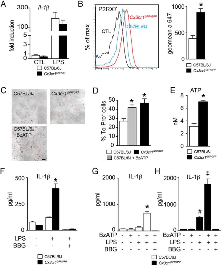
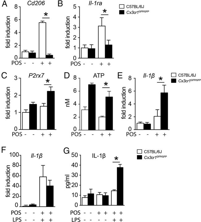
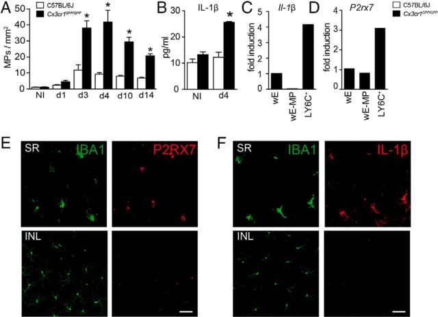
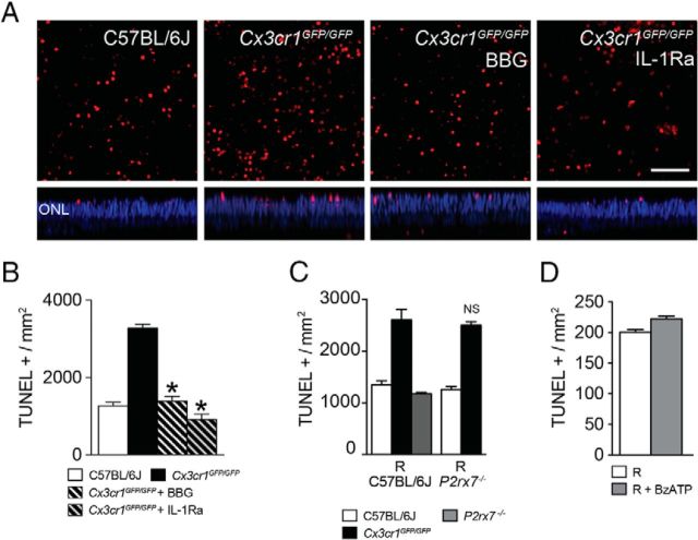
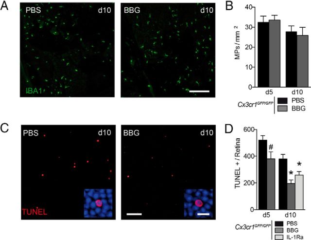
Photoreceptor degeneration in age-related macular degeneration (AMD) is associated with an infiltration and chronic accumulation of mononuclear phagocytes (MPs). We have previously shown that Cx3cr1-deficient mice develop age- and stress- related subretinal accumulation of MPs, which is associated with photoreceptor degeneration. Cx3cr1-deficient MPs have been shown to increase neuronal apoptosis through IL-1β in neuroinflammation of the brain. The reason for increased IL-1β secretion from Cx3cr1-deficient MPs, and whether IL-1β is responsible for increased photoreceptor apoptosis in Cx3cr1-deficient mice, has not been elucidated. Here we show that Cx3cr1-deficient MPs express increased surface P2X7 receptor (P2RX7), which stimulates IL-1β maturation and secretion. P2RX7 and IL-1β inhibition efficiently blunted Cx3cr1-MP-dependent photoreceptor apoptosis in a monocyte/retina coculture system and in light-induced subretinal inflammation of Cx3cr1-deficient mice in vivo. Our results provide an explanation for increased CX3CR1-dependent IL-1β secretion and suggest that IL-1β or P2RX7 inhibition can help inhibit the inflammation-associated photoreceptor cell loss in late AMD, including geographic atrophy, for which no efficient treatment currently exists.

Keywords: IL-1; P2RX7; inflammasome; monocytes; retina.


Figures

Figure 1.
Figure 1.
Cx3cr1GFP/GFP-BMMs constitutively activate their P2RX7 receptors and secrete IL-1β after TLR activation by LPS. A, Quantitative RT-PCR of Il-1β mRNA normalized with Rps26 mRNA of C57BL/6J- and Cx3cr1GFP/GFP-BMMs cultured for 18 h in control conditions or treated with LPS (n = 4 per group, one-way ANOVA followed by Bonferroni's post-test, no statistical differences between genotype; representative of 3 independent experiments). B, P2RX7 surface expression on CD11b+ LY6Chigh C57BL/6J- and Cx3cr1GFP/GFP-BMMs cultured for 18 h without LPS was analyzed by flow cytometry (left). The magnitude of P2RX7 surface expression was measured by the geometric mean fluorescence intensity of C57BL/6J- and Cx3cr1GFP/GFP-BMMs (right) (n = 3 per group, *p < 0.05; Mann–Whitney U test, representative of 3 independent experiments). C, Representative images of C57BL/6J- and Cx3cr1GFP/GFP-BMMs incubated for 10 min with To-pro-3 in the absence (C57BL/6J and Cx3cr1GFP/GFP) or presence of BzATP (C57BL/6J + BzATP). Scale bar, 50 μm. D, Quantification of the number of To-pro-3-positive Cx3cr1GFP/GFP-BMMs and To-pro-3-positive C57BL/6J-BMMs incubated in the absence (C57BL/6J) or presence of BzATP (C57BL/6J + BzATP) for 10 min (n = 5 per group, *p < 0.05; one-way ANOVA followed by Dunnett's post-test; C57BL/6J-BMMs as control, representative of 2 independent experiments). E, Quantification of the extracellular ATP release in the supernatant of C57BL/6J- and Cx3cr1GFP/GFP-BMMs cultivated for 18 h without LPS (n = 6 per group, *p < 0.05; Mann–Whitney U test). F, Quantification of IL-1β in the supernatant of C57BL/6J- and Cx3cr1GFP/GFP-BMMs in control conditions or primed with LPS (60 ng/ml) or LPS + BBG (60 ng/ml + 10 μm; n = 3, *p < 0.05; one-way ANOVA followed by Bonferroni's post-test, representative of 3 independent experiments). G, H, Quantification of IL-1β in the supernatant of C57BL/6J-BMMs (G) and Cx3cr1GFP/GFP-BMMs (H) primed overnight with LPS (60 ng/ml) and then stimulated with BzATP (1 mm) in the presence or the absence of BBG (10 μm; n = 5, *p < 0.05; one-way ANOVA followed by Bonferroni's post-test, representative of 2 independent experiments). CTL, control.
Figure 2.
Figure 2.
BMM differentiation in the presence of POSs. A–C, Quantitative RT-PCR of Cd206 (A), Il-1Ra (B), and P2rx7 (C) mRNA normalized with Rps26 mRNA of C57BL/6J- and Cx3cr1GFP/GFP-BMMs cultured for 18 h with or without POSs (n = 5 per group, *p < 0.01; one-way ANOVA followed by Bonferroni's post-test, representative of 3 independent experiments). D, Quantification of ATP release from C57BL/6J- and Cx3cr1GFP/GFP-BMMs cultivated for 18 h with or without POSs (n = 6 per group, *p < 0.01; one-way ANOVA followed by Bonferroni's post-test, representative of 3 independent experiments). E, Quantitative RT-PCR of Il-1β mRNA normalized with Rps26 mRNA of C57BL/6J- and Cx3cr1GFP/GFP-BMMs cultured for 18 h with or without POSs (n = 4 per group, *p < 0.01; one-way ANOVA followed by Bonferroni's post-test). F, Quantitative RT-PCR of Il-1β mRNA normalized with Rps26 mRNA of C57BL/6J- and Cx3cr1GFP/GFP-BMMs cultured for 18 h with or without POSs and LPS (60 ng/ml; n = 4 per group). G, Quantification by ELISA of IL-1β in the supernatants of C57BL/6J- and Cx3cr1GFP/GFP-BMMs cultured for 18 h with or without POSs and stimulated or not with LPS (60 ng/ml) for 2 h (n = 4 per group, *p < 0.01; one-way ANOVA followed by Bonferroni's post-test).
Figure 3.
Figure 3.
Subretinal MPs in light-challenged Cx3cr1GFP/GFP mice express IL-1β and P2RX7. A, IBA1-positive cell density in the subretinal space of C57BL/6J and Cx3cr1GFP/GFP mice at different time points during the light-challenge model (n = 5 per group, *p < 0.05; one-way ANOVA followed by Bonferroni's post-test). B, Quantification by ELISA of IL-1β protein in retinal protein extracts from PBS-perfused C57BL/6J and Cx3cr1GFP/GFP mice after 4 d of light-induced subretinal inflammation (n = 3 per group, *p < 0.01; one-way ANOVA followed by Bonferroni's post-test). C, Quantitative RT-PCR of Il-1β mRNA normalized with Rps26 of wE (set as 1), wE-MP, and FAC-sorted Ly6Chigh cells (LY6C+). (Each sample is a pool of cells from 8 independent eyes of PBS-perfused Cx3cr1GFP/GFP mice after 4 d of light challenge, values are the mean of two independent qPCR.) D, Quantitative RT-PCR of P2rx7 mRNA normalized with Rps26 of wE (set as 1), wE-MP, and FAC-sorted Ly6Chigh cells (LY6C+). (Each sample is a pool of cells from 8 independent eyes of PBS-perfused Cx3cr1GFP/GFP mice after 4 d of light challenge, values are the mean of two independent qPCR.) E, Costaining of P2RX7 (red) and IBA1 (green) in the SR (retinal flat mount, top) or INL (bottom) of Cx3cr1GFP/GFP mice after 4 d of light-induced subretinal inflammation. F, Costaining of IL-1β (red) and IBA1 (green) in the SR (choroidal/RPE flat mount, top) or INL (bottom) of Cx3cr1GFP/GFP mice after 4 d of light-induced subretinal inflammation. Scale bar, 50 μm. NI, non-illuminated; SR, subretinal space; INL, inner nuclear layer.
Figure 4.
Figure 4.
A P2RX7 antagonist and IL-1Ra inhibit Cx3cr1GFP/GFP-BMM photoreceptor toxicity. A, Confocal microscopy of TUNEL (red)-stained retinal flat mounts cultured for 18 h in contact with C57BL/6J-, Cx3cr1GFP/GFP-, and Cx3cr1GFP/GFP-BMMs in the presence of BBG (10 μm) or IL-1Ra (10 mg/ml). Nuclei were stained with Hoechst (blue). Scale bar, 50 μm. B, Quantification of TUNEL-positive nuclei/mm2 in the ONL of retinal flat mounts in contact with C57BL/6J-, Cx3cr1GFP/GFP-, and Cx3cr1GFP/GFP-BMMs in the presence or not of BBG (10 μm) or IL-1Ra (10 mg/ml; n = 4 per group, *p < 0.01; one-way ANOVA followed by Dunnett's post-test vs Cx3cr1GFP/GFP-BMMs, representative of at least 3 experiments). C, Quantification of TUNEL-positive nuclei/mm2 in the ONL of C57BL/6J- or P2rx7−/− explant with C57BL/6J-, Cx3cr1GFP/GFP, or P2rx7−/− BMMs (n = 4 per group, one-way ANOVA followed by Bonferroni's post-test; NS, no significant differences between C57BL/6J and P2rx7−/− explant cultured with Cx3cr1GFP/GFP-BMMs). D, Quantification of TUNEL-positive nuclei/mm2 in the ONL of retinal explant treated or not with 100 μm BzATP (n = 4 per experimental group, Mann–Whitney U test, no statistical difference was found). R, retina.
Figure 5.
Figure 5.
BBG and IL-1Ra inhibit subretinal inflammation-associated photoreceptor degeneration in Cx3cr1GFP/GFP mice. A, Representative accumulation of subretinal MP cells (choroidal/RPE flat mounts; green) at day 10 in PBS and BBG (25 mg/L) of Cx3cr1GFP/GFP mice treated by intravitreal (IVT) injections at days 3 and 7. Scale bar, 200 μm. B, MP density in the subretinal space of the light-challenge model of Cx3cr1GFP/GFP mice at days 5 and 10 treated with PBS and BBG at day 3 (animals kept until day 10 received a second IVT on day 7). (n = 6 per group, one-way ANOVA followed by Bonferroni's post-test, no differences were found between Cx3cr1GFP/GFP treated with PBS and BBG). C, Confocal microscopy of TUNEL-positive cells (red) through the ONL of a retinal flat mount at day 10 of the light-challenge model of Cx3cr1GFP/GFP mice treated with PBS or BBG (25 mg/L) by IVT injections. Scale bars: 50 μm, Inset, 10 μm. D, Quantification of the number of TUNEL-positive cell per retina of the light-challenge model of Cx3cr1GFP/GFP mice at days 5 and 10 treated with PBS, BBG (25 mg/ml), and IL-1Ra (150 mg/ml) at day 3 (animals kept until day 10 received a second IVT on day 7). (n = 5 per group, #p < 0.05; Mann–Whitney U test day 5 PBS vs day 5 BBG, *p < 0.05; one-way ANOVA followed by Dunnett's post-tests vs day 10 PBS).

Similar articles

Cited by

References

    1. Allan SM, Tyrrell PJ, Rothwell NJ. Interleukin-1 and neuronal injury. Nat Rev Immunol. 2005;5:629–640. doi: 10.1038/nri1664. - DOI - PubMed
    1. Arbeloa J, Pérez-Samartín A, Gottlieb M, Matute C. P2X7 receptor blockade prevents ATP excitotoxicity in neurons and reduces brain damage after ischemia. Neurobiol Dis. 2012;45:954–961. doi: 10.1016/j.nbd.2011.12.014. - DOI - PubMed
    1. Bartlett R, Yerbury JJ, Sluyter R. P2X7 receptor activation induces reactive oxygen species formation and cell death in murine EOC13 microglia. Mediators Inflamm. 2013;2013:271813. doi: 10.1155/2013/271813. - DOI - PMC - PubMed
    1. Bond SR, Naus CC. The pannexins: past and present. Front Physiol. 2014;5:58. doi: 10.3389/fphys.2014.00058. - DOI - PMC - PubMed
    1. Brändle U, Kohler K, Wheeler-Schilling TH. Expression of the P2X7-receptor subunit in neurons of the rat retina. Brain Res Mol Brain Res. 1998;62:106–109. doi: 10.1016/S0169-328X(98)00254-X. - DOI - PubMed

Publication types

MeSH terms