. 2015 May 6;35(18):7041-55.
doi: 10.1523/JNEUROSCI.5128-14.2015.

Distinct brainstem and forebrain circuits receiving tracheal sensory neuron inputs revealed using a novel conditional anterograde transsynaptic viral tracing system

Affiliations

Distinct brainstem and forebrain circuits receiving tracheal sensory neuron inputs revealed using a novel conditional anterograde transsynaptic viral tracing system

Alice E McGovern et al. J Neurosci. .

Abstract

Sensory nerves innervating the mucosa of the airways monitor the local environment for the presence of irritant stimuli and, when activated, provide input to the nucleus of the solitary tract (Sol) and paratrigeminal nucleus (Pa5) in the medulla to drive a variety of protective behaviors. Accompanying these behaviors are perceivable sensations that, particularly for stimuli in the proximal end of the airways, can be discrete and localizable. Airway sensations likely reflect the ascending airway sensory circuitry relayed via the Sol and Pa5, which terminates broadly throughout the CNS. However, the relative contribution of the Sol and Pa5 to these ascending pathways is not known. In the present study, we developed and characterized a novel conditional anterograde transneuronal viral tracing system based on the H129 strain of herpes simplex virus 1 and used this system in rats along with conventional neuroanatomical tracing with cholera toxin B to identify subcircuits in the brainstem and forebrain that are in receipt of relayed airway sensory inputs via the Sol and Pa5. We show that both the Pa5 and proximal airways disproportionately receive afferent terminals arising from the jugular (rather than nodose) vagal ganglia and the output of the Pa5 is predominately directed toward the ventrobasal thalamus. We propose the existence of a somatosensory-like pathway from the proximal airways involving jugular ganglia afferents, the Pa5, and the somatosensory thalamus and suggest that this pathway forms the anatomical framework for sensations arising from the proximal airway mucosa.

Keywords: HSV-1 H129; airway sensations; neuroanatomical tracing; nucleus of the solitary tract; paratrigeminal; visceral sensation.


Figures

Figure 1.
Figure 1.
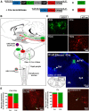
Conditional anterograde transneuronal viral tracing with H129floxed. A, H129floxed contains an expression cassette that enables the virus to switch reporters from EGFP to tdTomato in the presence of Cre-recombinase (Cre). B, Expression of Cre in discrete populations of central neurons is achieved by microinjection of an AAV-Cre expression vector driven by a HCMV promoter. In the example shown, AAV-Cre is delivered to the Sol and not the Pa5. Two weeks after AAV-Cre injection, the trachea is inoculated with H129floxed and, as the virus moves from airway primary sensory neurons in the vagal ganglia to infect medullary relay nuclei, it is recombined in the Sol, but not the Pa5, resulting in viral-induced expression of tdTomato in Sol neurons as well as all synaptically connected neurons in the circuits arising from the Sol. Neural circuits arising from neurons that do not express Cre (e.g., the Pa5) are also virally infected but display EGFP rather than tdTomato fluorescence. C, Infection of wild-type (−Cre) 3T3 cells with the H129floxed virus results in the exclusive expression of EGFP (top), whereas infection of 3T3 Cre-expressing cells (+Cre) results in the exclusive expression of tdTomato (bottom), indicating efficient and complete recombination of H129floxed. D, H129floxed retains normal anterograde transsynaptic motility and, in the absence of Cre expression in the CNS, airway inoculation with H129floxed results in a time-dependent appearance of EGFP expression (but not tdTomato) throughout the neuraxis (shown is EGFP expression in both the Sol and Pa5). E, Graphs showing examples of the quantification of EGFP- (green bars) and tdTomato (red bars)-expressing cells in the caudal and rostral ventrolateral medulla (CVL/ RVL) and paraventricular nucleus (Pa). Notably, the ratio of EGFP to tdTomato at these central sites differs depending on whether AAV-Cre is delivered to the Sol or Pa5, indicative of the relative contribution of the Sol and Pa5 to the projections that terminate in the CVL/RVL and Pa. Photomicrographs represent examples of CVL/RVL and Pa neurons expressing tdTomato versus EGFP in animals transduced with AAV-Cre into the Sol. Arrowheads show dual infected neurons (yellow), which were rare.
Figure 2.
Figure 2.
Differential terminations of jugular and nodose vagal ganglia neurons in the Pa5 and Sol. A, Example photomicrograph and corresponding line drawing of a caudal brainstem section showing the Sol and Pa5 locations for microinjection of CT-B tracers conjugated with 488 (green) or 594 (red) fluorophores, respectively. B, Bright-field photomicrograph of a whole-mount preparation of the rat vagal ganglia demonstrating the location of the jugular (JG) and nodose ganglia (NG) within the vagal sensory ganglia complex. C, Fluorescent photomicrograph of a whole-mount preparation of the rat vagal ganglia showing neuronal soma retrogradely labeled with CT-B from the Sol (green) and Pa5 (red). D, Quantitative cell counts performed on serial ganglia sections demonstrating that Pa5-projecting neurons reside in the jugular ganglia, whereas Sol projecting neurons are located within the nodose ganglia. Data are presented as the mean ± SEM; n = 7 dual tracing experiments. 9, Glossopharyngeal nerve; 10, vagus nerve; 11, accessory nerve; BS, brainstem; IO, inferior olives; PG, petrosal ganglia; Ph, pharyngeal branch; SLN, superior laryngeal nerve; Sp5, spinal trigeminal nucleus; sp5, spinal trigeminal tract.
Figure 3.
Figure 3.
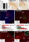
Differential medullary projections arising from the Sol and Pa5. A, Mean data showing quantification of the number of Cre-immunoreactive cells in serial brainstem sections of animals receiving microinjections of AAV-Cre into the Sol (n = 11 animals) versus Pa5 (n = 11 animals). Note the remarkably consistent number of Cre-expressing cells between the two groups. Representative photomicrographs show immunoperoxidase staining of Cre-expressing cells in the Sol (A′) and Pa5 (A′′). B, Representative photomicrograph showing many tdTomato-expressing neurons in the rostral ventrolateral medulla (RVL) after tracheal inoculation with H129floxed in animals expressing Cre in the Sol. B′, Representative photomicrograph of a matching brainstem section from an animal with CT-B488 and CT-B594 injected into the Sol and Pa5, respectively. Note the disproportionately greater density of direct monosynaptic projections from the Sol to the RVL. The graphs show the ratio of H129-infected tdTomato-expressing neurons in medullary nuclei of animals expressing Cre in the Sol (solid red bars) versus Pa5 (shaded red bars; C) and (C′) the ratio of CT-B488 (green bars, Sol injection) and CT-B594 (red bars, Pa5 injection)-labeled terminals in the same medullary nuclei shown in C. In each instance, the bars represent the ratio of the summed data obtained from six animals (viral tracing, C) and four animals (CT-B tracing, C′) and highlights differences in the relative contribution of inputs from the Sol and Pa5 to regions of interest. Mean data are presented in Tables 1 and 2. D, Representative photomicrograph of H129 virally infected tdTomato-expressing neurons in the Sol (arrows) in an animal transduced with Cre in the Pa5, indicative that some (albeit relatively few) airway afferent inputs to the Pa5 are relayed to the Sol. D′, Representative photomicrograph confirming extensive terminal fiber labeling in the Sol following CT-B594 injection into the Pa5. The inset (D′′) shows retrogradely labeled neurons in the Pa5 after microinjection of CT-B488 into the Sol. Comparable reciprocal connectivity was not observed. cc, Central canal; CVL, caudal ventrolateral medulla; IRt, intermediate reticular formation; 4V, fourth ventricle.
Figure 4.
Figure 4.
Differential pontine projections arising from the Sol and Pa5. A, Representative photomicrograph showing many tdTomato-expressing neurons in the parabrachial complex (particularly in the lateral parabrachial nucleus; LPB) after tracheal inoculation with H129floxed in animals expressing Cre in the Sol. A′, Representative photomicrograph of a matching brainstem section from an animal with CT-B488 and CT-B594 injected into the Sol and Pa5, respectively. Note the comparable distribution of direct monosynaptic projections from the Sol to the LPB compared with the Pa5 projections that are predominately in the medial parabrachial externus nucleus (MPBE). B, Representative photomicrograph of tdTomato-expressing neurons in the locus ceruleus (LC) after tracheal inoculation with H129floxed in animals expressing Cre in the Sol. The graphs show the ratio of H129-infected tdTomato-expressing neurons in pontine nuclei of animals expressing Cre in the Sol (solid red bars) versus Pa5 (shaded red bars) (C) and the ratio of CT-B488- (green bars, Sol injection) and CT-B594 (red bars, Pa5 injection)-labeled terminals (C′) in the same pontine nuclei shown in C. In each instance, the bars represent the ratio of the summed data obtained from six animals (viral tracing, C) and four animals (CT-B tracing, C′) and highlights differences in the relative contribution of inputs from the Sol and Pa5 to regions of interest. Mean data are presented in Tables 1 and 2. KF, Kölliker–Fuse; scp, superior cerebellar peduncle.
Figure 5.
Figure 5.
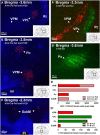
Differential hypothalamic, subthalamic, and amygdaloid projections arising from the Sol and Pa5. A, Representative photomicrograph showing many tdTomato-expressing neurons in the zona incerta (ZI) after tracheal inoculation with H129floxed in animals expressing Cre in the Sol. A′, Representative photomicrograph of a matching brainstem section from an animal with CT-B488 and CT-B594 injected into the Sol and Pa5, respectively. Note the comparable distribution of direct monosynaptic projections from the Sol to the ZI. B, C, Representative photomicrographs of tdTomato-expressing neurons in the central amygdala (CeA) and paraventricular nucleus of the hypothalamus (Pa) after tracheal inoculation with H129floxed in animals expressing Cre in the Sol. The graphs show the ratio of H129 infected tdTomato-expressing neurons in hypothalamic, subthalamic, and amygdaloid nuclei of animals expressing Cre in the Sol (solid red bars) versus Pa5 (shaded red bars) (D) and the ratio of CT-B488 (green bars, Sol injection) and CT-B594 (red bars, Pa5 injection) labeled terminals in the same nuclei (D′). In each instance, the bars represent the ratio of the summed data obtained from six animals (viral tracing, D) and four animals (CT-B tracing, D′) and highlights differences in the relative contribution of inputs from the Sol and Pa5 to regions of interest. Mean data are presented in Tables 1 and 2. BLA, Basolateral amygdala; ic, internal capsule; ml, medial lemniscus; SubT, subthalamic nucleus; VPL, ventral posterolateral thalamus; 3V, third ventricle.
Figure 6.
Figure 6.
Differential thalamic projections arising from the Sol and Pa5. A, Representative photomicrograph showing many tdTomato-expressing neurons in the ventral posterolateral nucleus (VPL) after tracheal inoculation with H129floxed in animals expressing Cre in the Pa5. Note the comparably fewer tdTomato-expressing neurons in the ventral posteromedial nucleus (VPM) of the same animal. A′, Representative photomicrograph of a matching brainstem section from an animal with CT-B488 and CT-B594 injected into the Sol and Pa5, respectively. Note the dichotomy of the distribution of direct monosynaptic projections from the Sol to the VPL and the Pa5 to the VPM, suggesting that the VPL neurons infected with H129floxed virus in A must receive indirect inputs from the Pa5. B, Representative photomicrograph showing many tdTomato-expressing neurons in the VPM and posterior group (Po) after tracheal inoculation with H129floxed in animals expressing Cre in the Sol. B′, Representative photomicrograph of labeled terminals in the Po after microinjection of CT-B488 into the Sol. C, Representative photomicrograph of tdTomato-expressing neurons in the submedius thalamus (SubM) after tracheal inoculation with H129floxed in animals expressing Cre in the Pa5. The graphs show the ratio of H129 infected tdTomato-expressing neurons in thalamic nuclei of animals expressing Cre in the Sol (solid red bars) versus Pa5 (shaded red bars) (D) and the ratio of CT-B488- (green bars, Sol injection) and CT-B594 (red bars, Pa5 injection)-labeled terminals (D′) in the same nuclei shown in C. In each instance, the bars represent the ratio of the summed data obtained from six animals (viral tracing, D) and four animals (CT-B tracing, D′) and highlights differences in the relative contribution of inputs from the Sol and Pa5 to regions of interest. Note the relatively few direct monosynaptic inputs (CT-B-labeled terminals) into the SubM (D′) from the Pa5. Mean data are presented in Tables 1 and 2. ml, Medial lemniscus; Rt, reticular nucleus; 3V, third ventricle.
Figure 7.
Figure 7.
Sagittal schematic of the rat brain summarizing the proposed differences in the airway sensory circuitry relayed via Sol and Pa5. Dashed and solid lines show inputs to nuclei predominately arising from Sol and Pa5 neurons, respectively. The appearance of infected neurons in the caudal and rostral ventral lateral medulla (CVL/RVL), intermediate reticular nucleus (IRt), spinal trigeminal (Sp5), lateral parabrachial (LPB), locus ceruleus (LC), zona incerta (ZI), paraventricular hypothalamus (Pa), lateral hypothalamus (LH), and central amygdala (CeA) occurred at earlier time points (48–96 h), suggestive of direct monosynaptic inputs from the Sol or Pa5. In contrast, viral infection in posterior (Po), submedius (SubM), ventral posterolateral (VPL), and ventral posteromedial (VPM) of the thalamus did not occur until the middle time points (96–120 h), indicative of a polysynaptic connection from the Sol or Pa5. Conventional tracing with CT-B further supports this notion.

References

    1. Agster KL, Burwell RD. Cortical efferents of the perirhinal, postrhinal, and entorhinal cortices of the rat. Hippocampus. 2009;19:1159–1186. doi: 10.1002/hipo.20578. - DOI - PMC - PubMed
    1. Alioto OE, Lindsey CJ, Koepp J, Caous CA. Sensory sciatic nerve afferent inputs to the dorsal lateral medulla in the rat. Auton Neurosci. 2008;140:80–87. doi: 10.1016/j.autneu.2008.04.006. - DOI - PubMed
    1. Arvidsson J, Thomander L. An HRP study of the central course of sensory intermediate and vagal fibers in peripheral facial nerve branches in the cat. J Comp Neurol. 1984;223:35–45. doi: 10.1002/cne.902230104. - DOI - PubMed
    1. Baker CV, Schlosser G. The evolutionary origin of neural crest and placodes. J Exp Zool B Mol Dev Evol. 2005;304:269–273. - PubMed
    1. Balan Júnior A, Caous CA, Yu YG, Lindsey CJ. Barosensitive neurons in the rat tractus solitarius and paratrigeminal nucleus: a new model for medullary, cardiovascular reflex regulation. Can J Physiol Pharmacol. 2004;82:474–484. doi: 10.1139/y04-054. - DOI - PubMed

Publication types

LinkOut - more resources