. 2015 May 6;35(18):7131-42.
doi: 10.1523/JNEUROSCI.5051-14.2015.

GIRK Channels Modulate Opioid-Induced Motor Activity in a Cell Type- and Subunit-Dependent Manner

Affiliations

GIRK Channels Modulate Opioid-Induced Motor Activity in a Cell Type- and Subunit-Dependent Manner

Lydia Kotecki et al. J Neurosci. .

Abstract

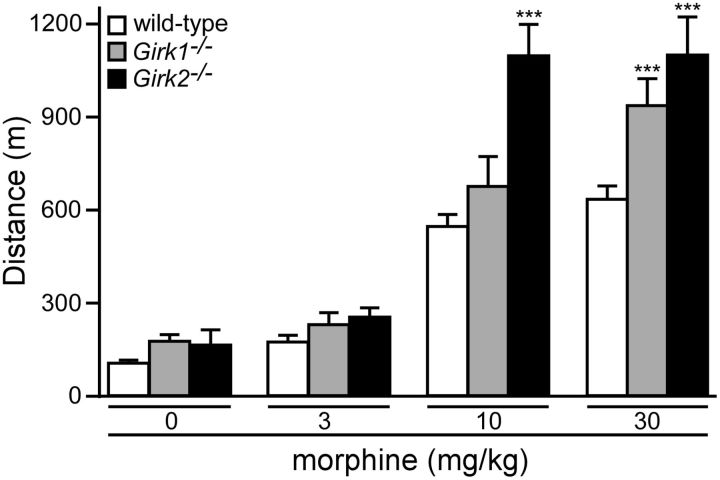
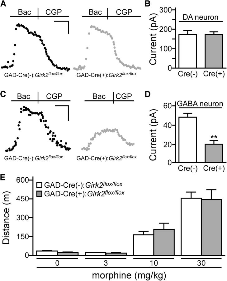
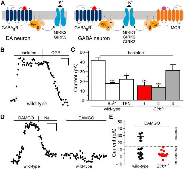
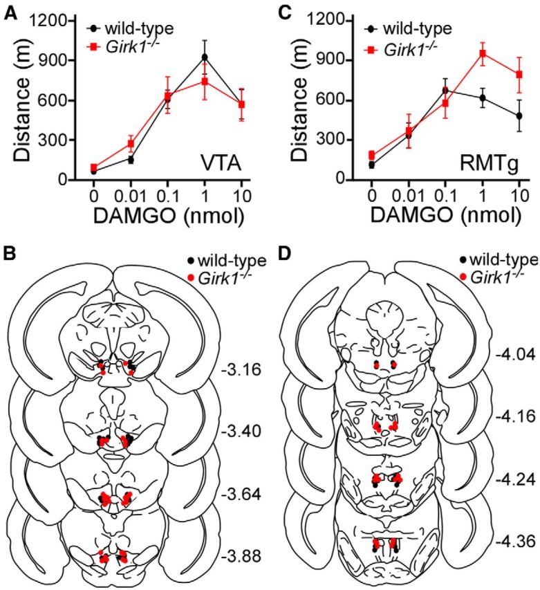
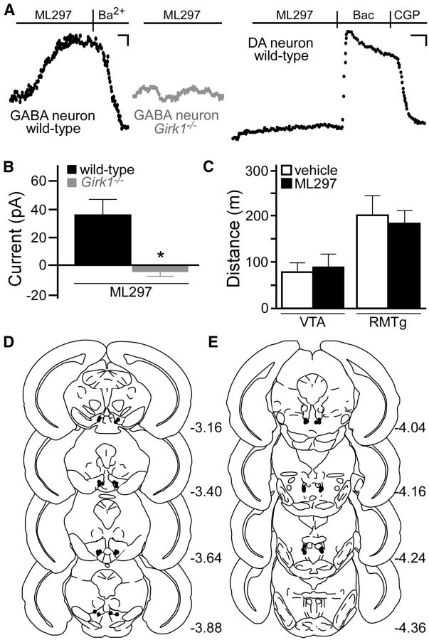
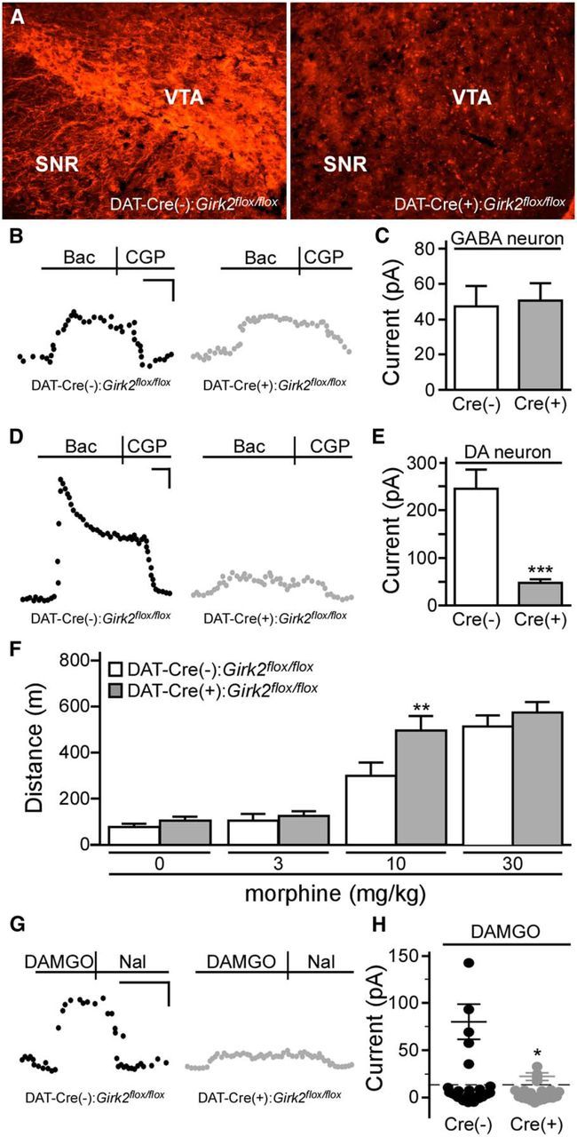
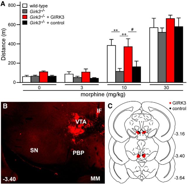
G-protein-gated inwardly rectifying K(+) (GIRK/Kir3) channel activation underlies key physiological effects of opioids, including analgesia and dependence. GIRK channel activation has also been implicated in the opioid-induced inhibition of midbrain GABA neurons and consequent disinhibition of dopamine (DA) neurons in the ventral tegmental area (VTA). Drug-induced disinhibition of VTA DA neurons has been linked to reward-related behaviors and underlies opioid-induced motor activation. Here, we demonstrate that mouse VTA GABA neurons express a GIRK channel formed by GIRK1 and GIRK2 subunits. Nevertheless, neither constitutive genetic ablation of Girk1 or Girk2, nor the selective ablation of GIRK channels in GABA neurons, diminished morphine-induced motor activity in mice. Moreover, direct activation of GIRK channels in midbrain GABA neurons did not enhance motor activity. In contrast, genetic manipulations that selectively enhanced or suppressed GIRK channel function in midbrain DA neurons correlated with decreased and increased sensitivity, respectively, to the motor-stimulatory effect of systemic morphine. Collectively, these data support the contention that the unique GIRK channel subtype in VTA DA neurons, the GIRK2/GIRK3 heteromer, regulates the sensitivity of the mouse mesolimbic DA system to drugs with addictive potential.

Keywords: GIRK; Kir3; conditional knockout; morphine; rostromedial tegmental area; ventral tegmental area.


Figures

Figure 1.
Figure 1.
Constitutive Girk subunit ablation and morphine-induced motor activity. The total distance traveled by adult male wild-type (white; n = 26/dose), Girk1−/− (gray; n = 18/dose), and Girk2−/− (black; n = 7/dose) mice during a 60 min test following systemic administration of saline (0) or morphine (3, 10, and 30 mg/kg, i.p.) is shown. Main effects of genotype (F(2,48) = 12.8, p < 0.0001) and dose (F(3,144) = 133.6, p < 0.0001) were observed, as well as a significant interaction between genotype and dose (F(6,144) = 5.4, p < 0.0001). ***p < 0.001 versus wild-type (within dose).
Figure 2.
Figure 2.
Generation of Girk2flox/flox mice. A, Depiction of the mouse Girk2/Kcnj6 gene, highlighting the seven known exons (rectangles with corresponding numbers below; Wei et al., 1998). Although several alternative splice variants have been identified, using two distinct translation initiation codons (ATG) and four distinct translation stop codons (TGA), all known variants contain exon 4. Shaded regions of exons denote protein-coding sequences. Some exons (1, 4, and 6) contain internal splice acceptor sites, denoted by vertical lines. B, Schematic depictions of a GIRK2 subunit. The image on the left shows the membrane topology of a GIRK2 subunit. Each subunit contains intracellular N- and C-terminal domains, two membrane-spanning domains (M1 and M2), two extracellular loops (×1 and ×2), and a pore domain (P). The schematic on the right is a linear depiction of the GIRK2a subunit, a prominent isoform in the brain (Wei et al., 1998). The region encoded by exon 4, which includes most of the N terminus (NT), both membrane-spanning domains and extracellular loops, the pore domain, and much of the C terminus (CT), is denoted by arrows. C, The Girk2 targeting strategy included engineering a loxP site just upstream (214 bp) of exon 4 and incorporating a NEO resistance cassette containing Flp recognition target (FRT) sites and a second loxP site; this cassette was positioned 795 bp downstream from exon 4. In total, the target region spanned 1.93 Kbp and was flanked by a 5′ homology arm (extending ∼5.4 Kbp upstream from exon 4) and a 3′ homology arm (an ∼7 Kbp fragment found immediately downstream from the target region). The NEO cassette was removed by crossing selected chimeras with C57BL/6N Flp deleter mice, yielding the Girk2flox allele. D, Tissue from the cortex (n = 9–10/genotype; t(17) = 0.09, p = 0.9), hippocampus (n = 6–7/genotype; t11 = 0.4, p = 0.7), and cerebellum (n = 7–8/genotype; t(13) = 1.3, p = 0.2) of wild-type and Girk2flox/flox (f/f) mice was evaluated for GIRK2 mRNA levels using quantitative RT-PCR. E, GIRK2 protein immunoreactivity (ir) relative to β-actin in samples from the cortex (n = 6–7/genotype; t(11) = 0.4, p = 0.7), hippocampus (n = 5–7/genotype; t(10) = 0.05, p = 1.0), and cerebellum (n = 4–7/genotype; t(9) = 0.6, p = 0.6) of wild-type and Girk2flox/flox (f/f) mice was assessed using quantitative immunoblotting. The level of GIRK2, which is seen as a doublet (top), was compared to the level of β-actin control (bottom) in each sample. Gel images of GIRK2 and β-actin were cropped and aligned with a molecular weight scale showing the markers for 52 and 38 kDA.
Figure 3.
Figure 3.
GABA neuron-specific ablation of GIRK channels and morphine-induced motor activity. A, Representative traces of currents evoked by baclofen (Bac; 200 μm) in putative VTA DA neurons from GAD-Cre(−):Girk2flox/flox (black) and GAD-Cre(+):Girk2flox/flox (gray) mice. Currents were reversed by the GABAB receptor antagonist CGP54626 (CGP; 2 μm). B, Summary plot of baclofen-induced currents in putative VTA DA neurons from GAD-Cre(−):Girk2flox/flox (white; n = 7) and GAD-Cre(+):Girk2flox/flox (gray; n = 8) mice. No genotype-dependent difference was observed (t(13) = 0.09, p = 0.9). C, Representative traces of currents evoked by baclofen (200 μm) in putative VTA GABA neurons from GAD-Cre(−):Girk2flox/flox (left, black) and GAD-Cre(+):Girk2flox/flox (right, gray) mice. D, Summary plot of baclofen-induced currents in putative VTA GABA neurons from GAD-Cre(−):Girk2flox/flox (white; n = 6) and GAD-Cre(+):Girk2flox/flox (gray; n = 5) mice. A significant genotype-dependent difference was observed (t(9) = 4.1, **p < 0.01). E, Total distance traveled (in meters) by GAD-Cre(−):Girk2flox/flox (white; n = 14) and GAD-Cre(+):Girk2flox/flox (gray; n = 7) mice during a 60 min period following systemic (intraperitoneal) administration of saline (0) or morphine (3, 10, and 30 mg/kg). A main effect of dose (F(3,57) = 76.6, p < 0.0001) was observed, but there was no main effect of genotype (F(1,19) = 0.02, p = 0.9), nor was there an interaction between genotype and dose (F(3,57) = 0.3, p = 0.8). Calibrations: A, 60 pA, 200 s; C, 20 pA, 200 s.
Figure 4.
Figure 4.
Characterization of the GIRK channel in VTA GABA neurons. A, Proposed GIRK subunit and receptor expression patterns in VTA DA and GABA neurons. B, Typical current induced by baclofen (200 μm) and reversed by CGP54626 (CGP; 2 μm) in a putative VTA GABA neuron from a wild-type mouse. C, The impact of pharmacologic and genetic ablation of GIRK channels on baclofen-induced currents in putative VTA GABA neurons (F(5,58) = 12.5; p < 0.0001). Currents are plotted for putative VTA GABA neurons from wild-type mice in the absence (control; n = 21) or presence of external Ba2+ (0.3 mm; n = 8) or TPN (200 nm; n = 4), and in neurons from Girk1−/− (red; n = 9), Girk2−/− (gray; n = 14), and Girk3−/− mice (gray; n = 8). *p < 0.05, ***p < 0.001 versus control. D, Responses of two VTA GABA neurons (eGFP-positive VTA neurons in slices from GAD67-eGFP mice) to a saturating concentration of DAMGO (3 μm). The response observed in the neuron shown on the left was reversed by naloxone (Nal; 10 μm). E, Summary of DAMGO-induced currents in putative VTA GABA neurons from wild-type (4 of 15 responders; 28.6 ± 2.7 pA) and Girk1−/− (0 of 13 responders) mice. Approximately one-third (4 of 15) of putative VTA GABA neurons evaluated exhibited a response to DAMGO (defined as a naloxone (Nal)-reversible current of >15 pA); none of the neurons in slices from Girk1−/− mice exhibited a response to DAMGO. Calibrations: B, D, 5 pA, 120 s.
Figure 5.
Figure 5.
Intracranial DAMGO-induced motor activity. A, Open-field motor activity of adult male wild-type (black; n = 15) and congenic Girk1−/− (red; n = 15) mice during a 60 min period following bilateral intra-VTA infusion of 0 nmol (saline; n = 15/genotype), 0.01 nmol (n = 5–6/genotype), 0.1 nmol (6–7/genotype), 1 nmol (5–8/genotype), and 10 nmol (5–8/genotype) of DAMGO. A main effect of DAMGO dose was observed (F(4,70) = 36.2, p < 0.001), but there was no effect of genotype (F(1,70) = 0.005, p = 1.0) or genotype/concentration interaction (F(4,70) = 0.8, p = 0.5). B, Schematic of coronal sections (from bregma) depicting cannulae placements in mice used for intra-VTA experiments. C, Open-field motor activity of male wild-type (black; n = 11) and congenic Girk1−/− (red; n = 10) mice during a 60 min test following bilateral intra-RMTg infusion of 0 nmol (saline; n = 10–11/genotype), 0.01 nmol (n = 5–7/genotype), 0.1 nmol (5–7/genotype), 1 nmol (6–10/genotype, and 10 nmol (6–10/genotype) of DAMGO. A main effect of DAMGO dose (F(4,67) = 18.2, p < 0.001) and genotype (F(1,67) = 5.2, p < 0.05) was observed, but there was no genotype/concentration interaction (F(4,67) = 2.0, p = 0.1). D, Schematic of cannulae placements in mice evaluated in the intra-RMTg DAMGO study.
Figure 6.
Figure 6.
Direct activation of GIRK1-containing GIRK channels and motor activity. A, ML297 (10 μm) evoked a Ba2+-sensitive (0.3 mm) outward current in putative VTA GABA neurons from wild-type (left, black) but not Girk1−/− (middle, gray) mice. ML297 did not evoke an outward current in VTA DA neurons (right, black; n = 6), as shown in this recording from an eGFP-positive neuron from a Pitx3-eGFP mouse. Calibrations: left and middle, 15 pA, 180 s; right, 30 pA, 200 s. B, Summary of ML297-induced currents in putative VTA GABA neurons from wild-type (n = 6) and Girk1−/− (n = 5) mice (t(9) = 2.9). *p < 0.05). C, Open-field motor activity of male wild-type mice during a 60 min test following bilateral infusion of vehicle (0.1% DMSO; white) or ML297 (10 nmol; black) into the VTA (n = 10/group, t(18) = 0.4, p = 0.7) or RMTg (n = 9/group, t(16) = 0.3, p = 0.7). D, E, Schematic of coronal sections (from bregma) depicting cannulae placements in mice used for the intra-VTA and intra-RMTg ML297 studies.
Figure 7.
Figure 7.
DA neuron-specific ablation of GIRK channels and systemic morphine-induced motor activity. A, GIRK2 immunolabeling in midbrain sections from DAT-Cre(−):Girk2flox/flox and DAT-Cre(+):Girk2flox/flox mice. Images shown were taken from slices −2.98 mm relative to bregma. SNR, Substantia nigra pars reticulata. B, Representative traces evoked by baclofen (Bac, 200 μm) in putative VTA GABA (eGFP-negative) neurons from Pitx3-eGFP(+)/DAT-Cre(−):Girk2flox/flox (left, black) and Pitx3-eGFP(+)/DAT-Cre(+):Girk2flox/flox (right, gray) mice. C, Summary of baclofen-induced currents in putative VTA GABA (eGFP-negative) neurons from Pitx3-eGFP(+)/DAT-Cre(−):Girk2flox/flox (white; n = 10) and Pitx3-eGFP(+)/GADCre(+):Girk2flox/flox (gray; n = 7) mice (t(15) = 0.2, p = 0.8). D, Representative currents evoked by baclofen (200 μm) in VTA DA (eGFP-positive) neurons from Pitx3-eGFP(+)/DAT-Cre(−):Girk2flox/flox (left, black) and Pitx3-eGFP(+)/DAT-Cre(+):Girk2flox/flox (right, gray) mice. E, Summary of baclofen-induced currents in VTA DA (eGFP-positive) neurons from Pitx3-eGFP(+)/DAT-Cre(−):Girk2flox/flox (white; n = 10) and Pitx3-eGFP(+)/DAT-Cre(+):Girk2flox/flox (gray; n = 12) mice (t(20) = 5.2, ***p < 0.0001). F, Open-field motor activity of DAT-Cre(−):Girk2flox/flox (white; n = 6/dose) and DAT-Cre(+):Girk2flox/flox (gray; n = 12/dose) mice during a 60 min period following systemic administration of saline (0) or morphine (3, 10, and 30 mg/kg). Main effects of dose (F(3,48) = 85.3, p < 0.0001) and genotype (F(1,16) = 2.9, p = 0.1) were observed, along with a dose/genotype interaction (F(3,48) = 2.9, p < 0.05). G, Responses of DA neurons from DAT-Cre(−):Girk2flox/flox and DAT-Cre(+):Girk2flox/flox mice to a saturating concentration of DAMGO (3 μm). DAMGO-induced currents were reversed by naloxone (Nal, 10 μm). H, Summary of DAMGO-induced currents in VTA DA neurons from DAT-Cre(−):Girk2flox/flox (5/25 responders; 80.1 ± 18.4 pA) and DAT-Cre(+):Girk2flox/flox (4/20 responders; 22.7 ± 3.4 pA) mice. Calibrations: B, G, 40 pA, 200 s; D, 60 pA, 200 s. *p < 0.05 versus DAT-Cre(−):Girk2flox/flox mice; **p < 0.01 versus DAT-Cre(−):Girk2flox/flox mice; ***p < 0.001 versus DAT-Cre(-):Girk2flox/flox mice.
Figure 8.
Figure 8.
Girk3 ablation and rescue: morphine-induced motor activity. A, Total distance traveled by wild-type (white; n = 6) and Girk3−/− (gray; n = 6) mice, as well as Girk3−/− mice given intra-VTA GIRK3 (red; n = 5) or control (black; n = 5) lentivirus, during a 60 min period following systemic administration of saline (0) or morphine (3, 10, and 30 mg/kg). A main effect of dose (F(3,54) = 103.4, p < 0.0001) and genotype (F(3,18) = 3.2, p < 0.05) was observed, but there was no dose/genotype interaction (F(9,54) = 1.8, p = 0.1). **p < 0.01 versus wild-type; ++p < 0.01 versus Girk3−/−; #p < 0.05 versus Girk3−/− + control. B, Representative image showing lentiviral-driven GIRK3-YFP (YFP expression shown in red) expression in the VTA of a Girk3−/− mouse. SN, Substantia nigra; PBP, parabrachial pigmented nucleus; IF, interfascicular nucleus; MM, medial mammillary nucleus. C, Schematic of coronal sections (from bregma) depicting viral expression in mice included in this study.

Similar articles

Cited by

References

    1. Arora D, Haluk DM, Kourrich S, Pravetoni M, Fernández-Alacid L, Nicolau JC, Luján R, Wickman K. Altered neurotransmission in the mesolimbic reward system of Girk mice. J Neurochem. 2010;114:1487–1497. doi: 10.1111/j.1471-4159.2010.06864.x. - DOI - PMC - PubMed
    1. Arora D, Hearing M, Haluk DM, Mirkovic K, Fajardo-Serrano A, Wessendorf MW, Watanabe M, Luján R, Wickman K. Acute cocaine exposure weakens GABA(B) receptor-dependent G-protein-gated inwardly rectifying K+ signaling in dopamine neurons of the ventral tegmental area. J Neurosci. 2011;31:12251–12257. doi: 10.1523/JNEUROSCI.0494-11.2011. - DOI - PMC - PubMed
    1. Bäckman CM, Malik N, Zhang Y, Shan L, Grinberg A, Hoffer BJ, Westphal H, Tomac AC. Characterization of a mouse strain expressing Cre recombinase from the 3′ untranslated region of the dopamine transporter locus. Genesis. 2006;44:383–390. doi: 10.1002/dvg.20228. - DOI - PubMed
    1. Barrot M, Sesack SR, Georges F, Pistis M, Hong S, Jhou TC. Braking dopamine systems: a new GABA master structure for mesolimbic and nigrostriatal functions. J Neurosci. 2012;32:14094–14101. doi: 10.1523/JNEUROSCI.3370-12.2012. - DOI - PMC - PubMed
    1. Beckstead MJ, Grandy DK, Wickman K, Williams JT. Vesicular dopamine release elicits an inhibitory postsynaptic current in midbrain dopamine neurons. Neuron. 2004;42:939–946. doi: 10.1016/j.neuron.2004.05.019. - DOI - PubMed

Publication types

MeSH terms

Substances

LinkOut - more resources