. 2015 May 6;35(18):7264-71.
doi: 10.1523/JNEUROSCI.3862-14.2015.

Endogenous opioid activity in the anterior cingulate cortex is required for relief of pain

Affiliations

Endogenous opioid activity in the anterior cingulate cortex is required for relief of pain

Edita Navratilova et al. J Neurosci. .

Abstract

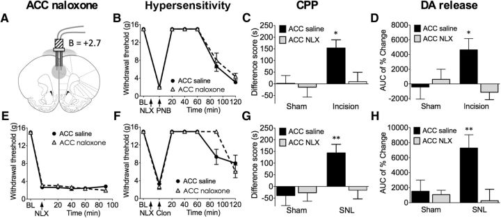
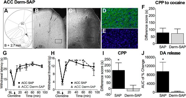
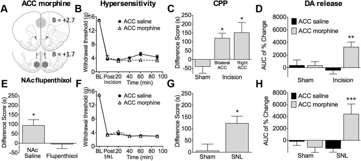
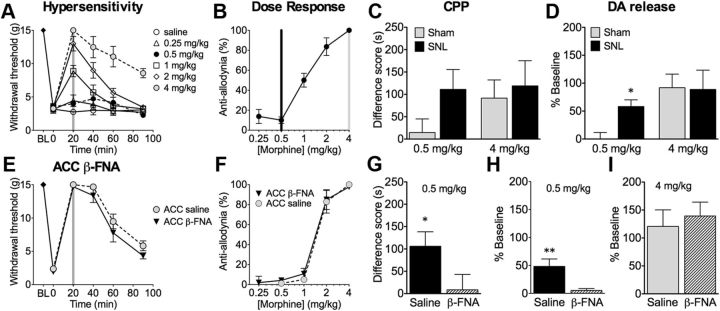
Pain is aversive, and its relief elicits reward mediated by dopaminergic signaling in the nucleus accumbens (NAc), a part of the mesolimbic reward motivation pathway. How the reward pathway is engaged by pain-relieving treatments is not known. Endogenous opioid signaling in the anterior cingulate cortex (ACC), an area encoding pain aversiveness, contributes to pain modulation. We examined whether endogenous ACC opioid neurotransmission is required for relief of pain and subsequent downstream activation of NAc dopamine signaling. Conditioned place preference (CPP) and in vivo microdialysis were used to assess negative reinforcement and NAc dopaminergic transmission. In rats with postsurgical or neuropathic pain, blockade of opioid signaling in the rostral ACC (rACC) inhibited CPP and NAc dopamine release resulting from non-opioid pain-relieving treatments, including peripheral nerve block or spinal clonidine, an α2-adrenergic agonist. Conversely, pharmacological activation of rACC opioid receptors of injured, but not pain-free, animals was sufficient to stimulate dopamine release in the NAc and produce CPP. In neuropathic, but not sham-operated, rats, systemic doses of morphine that did not affect withdrawal thresholds elicited CPP and NAc dopamine release, effects that were prevented by blockade of ACC opioid receptors. The data provide a neural explanation for the preferential effects of opioids on pain affect and demonstrate that engagement of NAc dopaminergic transmission by non-opioid pain-relieving treatments depends on upstream ACC opioid circuits. Endogenous opioid signaling in the ACC appears to be both necessary and sufficient for relief of pain aversiveness.

Keywords: affective dimension of pain; anterior cingulate cortex; neuropathic pain; nucleus accumbens; postsurgical pain; reward.


Figures

Figure 1.
Figure 1.
Blockade of opioid signaling in the rACC with naloxone prevents pain relief-induced CPP and NAc DA release in animals with incisional or neuropathic pain without altering evoked hypersensitivity. A, The rACC injection site for saline or naloxone (NLX; 3 μg, 10 min before testing). B, In rats with incisional injury, administration of saline or naloxone in the rACC had no effects on tactile hypersensitivity and its reversal by PNB with lidocaine injection into the popliteal fossa of the injured limb (n = 6). C, PNB produced significant preference only in incised rats pretreated with rACC saline but not naloxone (n = 13–23). D, In incised rats, rACC naloxone abolished PNB-induced NAc DA release (n = 5–10). E, In SNL rats, rACC saline or naloxone had no effect on tactile thresholds (n = 6–7). F, Neither treatment altered intrathecal clonidine-mediated reversal of tactile hypersensitivity (n = 6–7). G, Intrathecal clonidine produced significant preference only in SNL rats pretreated with rACC saline but not naloxone (n = 13–14). H, In SNL rats, rACC naloxone blocked intrathecal clonidine-induced DA release (n = 8–11). Two-way ANOVA with Bonferroni's comparison: *p < 0.05, **p < 0.01. Data are means ± SEMs.
Figure 2.
Figure 2.
Ablation of MOR-expressing neurons in the rACC with Derm-SAP prevents pain relief-induced CPP and DA release in injured rats with no effects on evoked hypersensitivity. A, Derm-SAP or blank SAP (1.5 pmol) was administered in the rACC 28–32 d before experimentation. Coronal brain sections at the level of the rACC were labeled with a rabbit polyclonal anti-MOR antibody. Micrographs demonstrate lack of MOR staining in the rACC of Derm-SAP-pretreated rats (C) but not in SAP animals (B). D and E show higher-magnification (20× objective) images of the rectangular areas outlined in B and C, respectively; MOR-positive neurons are in green, and DAPI nuclear staining is in blue. Images were acquired and processed using identical settings. F, Derm-SAP ablation does not interfere with cocaine-induced CPP, demonstrating that the deficit in MOR signaling in the rACC does not influence the animals' ability to learn and experience cocaine reward (n = 21–22). G, In SNL rats, rACC SAP or Derm-SAP did not block the development of thermal hyperalgesia nor its reversal by spinal clonidine (n = 5–6). H, SAP or Derm-SAP did not block tactile allodynia or its reversal by spinal clonidine (n = 5–9). I, Spinal clonidine produced significant preference in SAP-treated but not in Derm-SAP-treated SNL rats (n = 11–14). J, In SNL rats, ablation of MOR neurons abolished intrathecal clonidine-induced DA release (n = 8). Student's t test: *p < 0.05. Data are means ± SEMs.
Figure 3.
Figure 3.
Administration of morphine in the rACC relieves pain. A, The rACC injection site for morphine (20 μg/site) and the NAc injection site for α-flupenthixol (3 μg/site). B, In rats with postsurgical pain, bilateral administration of morphine (20 μg) in the rACC had no effect on tactile hyperalgesia (n = 7–8). C, Bilateral (20 μg) or contralateral (20 μg; right side) injections of morphine in the rACC produced CPP in incised rats (n = 11–39). D, Contralateral rACC morphine produced NAc DA release in incised rats (n = 8–15). F, Rats with SNL developed tactile hypersensitivity that was not reversed by bilateral rACC morphine (n = 12–17). G, In neuropathic but not sham rats, bilateral administration of morphine into the rACC produced CPP (n = 16–21). H, In neuropathic rats, bilateral morphine administration into the rACC elicited NAc DA release (n = 9–10). E, CPP induced by bilateral rACC morphine was abolished by pretreatment in the NAc with bilateral α-flupenthixol (3 μg, 10 min before; n = 15–16). *p < 0.05, **p < 0.01, ***p < 0.001. Data are means ± SEMs.
Figure 4.
Figure 4.
ACC β-FNA pretreatment blocks the effects of morphine on affective but not sensory aspects of pain. A, Intravenous administration of morphine time and dose dependently reversed tactile hypersensitivity (n = 5–7): 4.0 mg/kg morphine significantly attenuated tactile hypersensitivity at all time points (n = 5–7), whereas 0.5 mg/kg morphine had no significant effect on paw-withdrawal thresholds. B, The dose–response curve was calculated at 20 min after morphine. C, Morphine at 0.5 mg/kg produced CPP only in SNL but not sham rats; 4.0 mg/kg morphine produced CPP in both sham rats (n = 21–30). D, Morphine at 0.5 mg/kg elicited NAc DA release in SNL animals but not in sham-operated controls, whereas 4.0 mg/kg morphine produced NAc DA release in both sham and SNL animals (n = 10–11). E, SNL rats were pretreated 20–24 h before testing with bilateral β-FNA (3 μg) or saline in the rACC. Morphine at 4 mg/kg significantly elevated paw-withdrawal thresholds in both groups (n = 5–6). F, The dose–response curves were calculated at 20 min after morphine. G, In SNL animals pretreated with rACC saline, 0.5 mg/kg morphine elicited CPP, which was significantly attenuated in rACC β-FNA pretreated rats (n = 20–27). H, In SNL animals receiving 0.5 mg/kg morphine, pretreatment with rACC β-FNA, but not saline, abolished DA release (n = 11). I, In sham-operated rats morphine (4 mg/kg) mediated DA efflux was not affected by pretreatment with either saline or β-FNA (n = 7–9). *p < 0.05, **p < 0.01. Data are means ± SEMs.

Similar articles

Cited by

References

    1. Acworth I, Cunningham ML. The measurement of monoamine neurotransmitters in microdialysis perfusates using HPLC-ECD. Methods Mol Med. 1999;22:219–236. doi: 10.1385/0-89603-612-X:219. - DOI - PubMed
    1. Apkarian AV, Bushnell MC, Treede RD, Zubieta JK. Human brain mechanisms of pain perception and regulation in health and disease. Eur J Pain. 2005;9:463–484. doi: 10.1016/j.ejpain.2004.11.001. - DOI - PubMed
    1. Becerra L, Navratilova E, Porreca F, Borsook D. Analogous responses in the nucleus accumbens and cingulate cortex to pain onset (aversion) and offset (relief) in rats and humans. J Neurophysiol. 2013;110:1221–1226. doi: 10.1152/jn.00284.2013. - DOI - PMC - PubMed
    1. Becker S, Gandhi W, Schweinhardt P. Cerebral interactions of pain and reward and their relevance for chronic pain. Neurosci Lett. 2012;520:182–187. doi: 10.1016/j.neulet.2012.03.013. - DOI - PubMed
    1. Brennan TJ, Vandermeulen EP, Gebhart GF. Characterization of a rat model of incisional pain. Pain. 1996;64:493–501. doi: 10.1016/0304-3959(95)01441-1. - DOI - PubMed

Publication types