. 2015 May 6;35(18):7287-94.
doi: 10.1523/JNEUROSCI.5215-14.2015.

Layer 4 pyramidal neurons exhibit robust dendritic spine plasticity in vivo after input deprivation

Affiliations

Layer 4 pyramidal neurons exhibit robust dendritic spine plasticity in vivo after input deprivation

Amaya Miquelajauregui et al. J Neurosci. .

Abstract

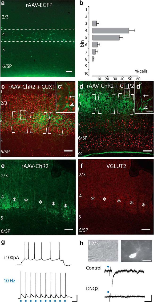
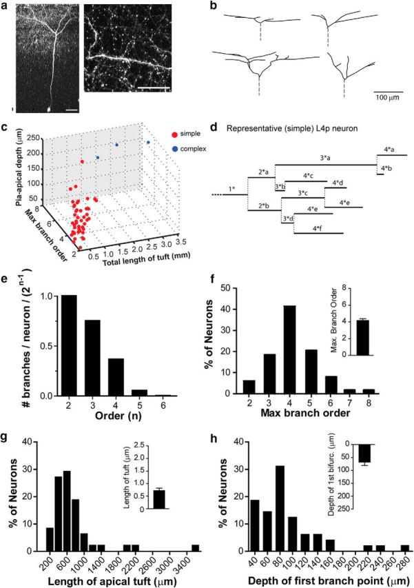
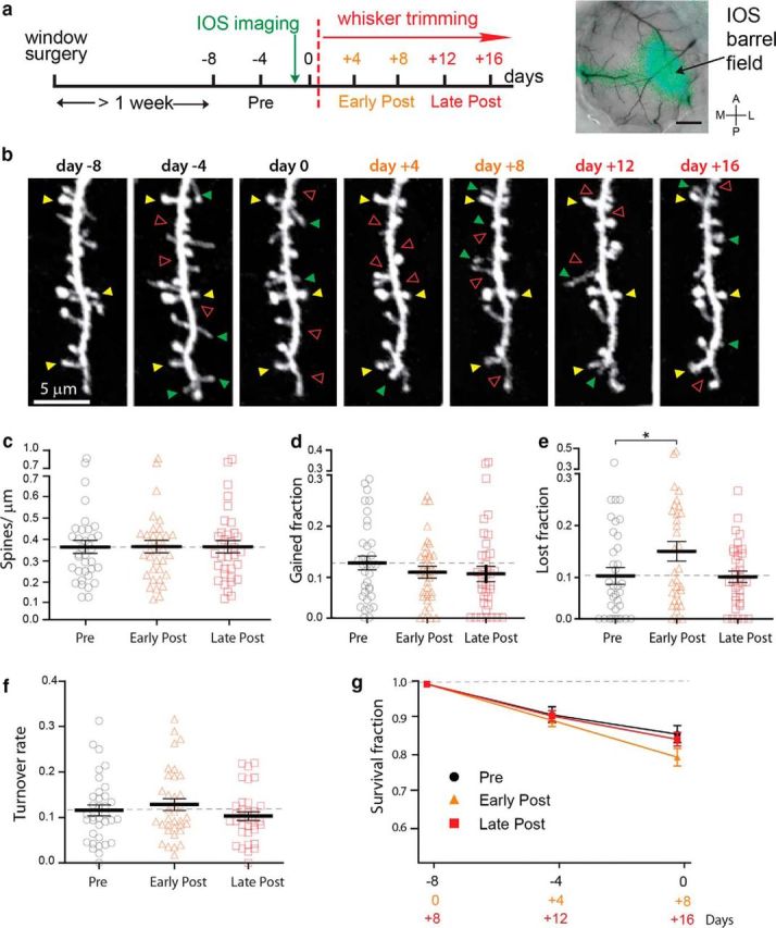
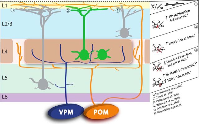
Pyramidal neurons in layers 2/3 and 5 of primary somatosensory cortex (S1) exhibit somewhat modest synaptic plasticity after whisker input deprivation. Whether neurons involved at earlier steps of sensory processing show more or less plasticity has not yet been examined. Here, we used longitudinal in vivo two-photon microscopy to investigate dendritic spine dynamics in apical tufts of GFP-expressing layer 4 (L4) pyramidal neurons of the vibrissal (barrel) S1 after unilateral whisker trimming. First, we characterize the molecular, anatomical, and electrophysiological properties of identified L4 neurons in Ebf2-Cre transgenic mice. Next, we show that input deprivation results in a substantial (∼50%) increase in the rate of dendritic spine loss, acutely (4-8 d) after whisker trimming. This robust synaptic plasticity in L4 suggests that primary thalamic recipient pyramidal neurons in S1 may be particularly sensitive to changes in sensory experience. Ebf2-Cre mice thus provide a useful tool for future assessment of initial steps of sensory processing in S1.

Keywords: Ebf2; barrel cortex; electrophysiology; optogenetics; two-photon; whisker.


Figures

Figure 1.
Figure 1.
Ebf2 labels L4 neurons in somatosensory cortex. a, Coronal section through S1 of an adult Ebf2-Cre mouse injected at E15 with rAAV-EGFP. b, Cell quantification of Ebf2+ cells across cortical layers. Dashed lines in a delineate L4 (bins 4–5). Approximately 84% of Ebf2+ cells in the neocortex of Ebf2-Cre;rAAV-EGFP mice were in L4. c, d, Adult coronal Ebf2-Cre;rAAV-ChR2 sections immunostained against Cux1 (c) and Ctip2 (d; both red) to label boundaries of L2–4 and L5–6, respectively. Insets (c′, d′) are confocal sections of representative pyramidal neurons (arrowheads) with a prominent apical dendrite (arrows). e, f, Ebf2-Cre;rAAV-ChR2 expression at P11 broadly overlaps with VGlut2+ expression (red) at individual barrels (asterisks). g, Representative whole-cell patch-clamp recordings in acute slices of an adult Ebf2+p neuron in response to a depolarizing current injection (100 pA; top) and to optogenetic stimulation (3 ms, 10 Hz; bottom). h, Optogenetic stimulation of Ebf2+ L4 neurons (3 ms, single pulse) triggers an EPSP in a L2/3 pyramidal neuron (middle) targeted under DIC optics (top left), and filled intracellularly with AlexaFluor 594 (top right). The response is completely blocked by DNQX (bottom). Scale bars: af, 100 μm; c′, d′, 50 μm; h (top), 20 μm; g, h, 20 mV and 100 ms.
Figure 2.
Figure 2.
Morphology of Ebf2+ pyramidal neurons in L4 (apical tufts). a, Two-photon image of a representative Ebf2+ L4 pyramidal neuron (soma depth ∼453 μm) and a dendritic segment in L1 (max proj, 17 slices, 2 μm apart) acquired in vivo in an adult Ebf2-Cre;rAAV-EGFP mouse. Scale bars: a, 50 μm; b, 100 μm. b, Neurolucida reconstructions of apical dendritic tufts from four representative neurons imaged in vivo. c, Ebf2+p neurons were segregated into two groups, simple (red) and complex (blue), using a k means test in MATLAB (see Materials and Methods) using values in f–h. d, Representative dendrogram of the L4 Ebf2+p apical tuft shown in a. e, Fraction of neurons with dendrites of a given order “n” (first order values, primary dendrites are excluded). fh, Frequency distribution histograms for maximum branch order (f), total length of the apical dendritic tuft (g), and depth of first bifurcation from the pial surface (h) for all reconstructed Ebf2+p neurons (n = 48). Insets show mean ± SEM.
Figure 3.
Figure 3.
Spine dynamics and experience-dependent plasticity of Ebf2+ L4 pyramidal neurons. a, Experimental design (left) and representative IOS map of barrel cortex (right). Scale bar: 100 μm. A, Anterior; P, posterior; M, medial; L, lateral. b, Example of longitudinal in vivo two-photon imaging of the same dendritic fragment before and after contralateral whisker trimming (pre or basal conditions: days −8,−4, and 0, black; early-post: days +4 and +8, orange; and late-post: days +12 and +16, red) from an adult Ebf2-Cre;rAAV-EGFP mouse. Images are best projections of ∼5–7 slices, 1 μm apart. Yellow, green, and red arrowheads indicate examples of persistent, gained, and lost spines, respectively. cf, Scatter plots of spine density (c), spine turnover rate (f), and fractions of gained (d) and lost (e) spines (mean ± SEM; *p < 0.05, linear mixed model). g, Survival fraction of spines over 4 and 8 d (during the pre, early-post, and late-post time intervals).
Figure 4.
Figure 4.
Experience-dependent spine plasticity in barrel cortex. Summary diagram of spine changes in apical tufts of neurons from different layers, documented by in vivo two-photon imaging studies. TOR, Turnover rate; FR, follicle removal.

Similar articles

Cited by

References

    1. Chen CC, Lu J, Zuo Y. Spatiotemporal dynamics of dendritic spines in the living brain. Front Neuroanat. 2014;8:28. doi: 10.3389/fnana.2014.00028. - DOI - PMC - PubMed
    1. Chiara F, Badaloni A, Croci L, Yeh ML, Cariboni A, Hoerder-Suabedissen A, Consalez GG, Eickholt B, Shimogori T, Parnavelas JG, Rakić S. Early B-cell factors 2 and 3 (EBF2/3) regulate early migration of Cajal-Retzius cells from the cortical hem. Dev Biol. 2012;365:277–289. doi: 10.1016/j.ydbio.2012.02.034. - DOI - PMC - PubMed
    1. Constantinople CM, Bruno RM. Deep cortical layers are activated directly by thalamus. Science. 2013;340:1591–1594. doi: 10.1126/science.1236425. - DOI - PMC - PubMed
    1. Cruz-Martín A, Crespo M, Portera-Cailliau C. Delayed stabilization of dendritic spines in fragile X mice. J Neurosci. 2010;30:7793–7803. doi: 10.1523/JNEUROSCI.0577-10.2010. - DOI - PMC - PubMed
    1. Feldmeyer D, Brecht M, Helmchen F, Petersen CC, Poulet JF, Staiger JF, Luhmann HJ, Schwarz C. Barrel cortex function. Prog Neurobiol. 2013;103:3–27. doi: 10.1016/j.pneurobio.2012.11.002. - DOI - PubMed

Publication types