Review
. 2015 Aug;72(16):3051-67.
doi: 10.1007/s00018-015-1914-2. Epub 2015 May 7.

Control of polarized assembly of actin filaments in cell motility

Affiliations
Review

Control of polarized assembly of actin filaments in cell motility

Marie-France Carlier et al. Cell Mol Life Sci. 2015 Aug.

Abstract

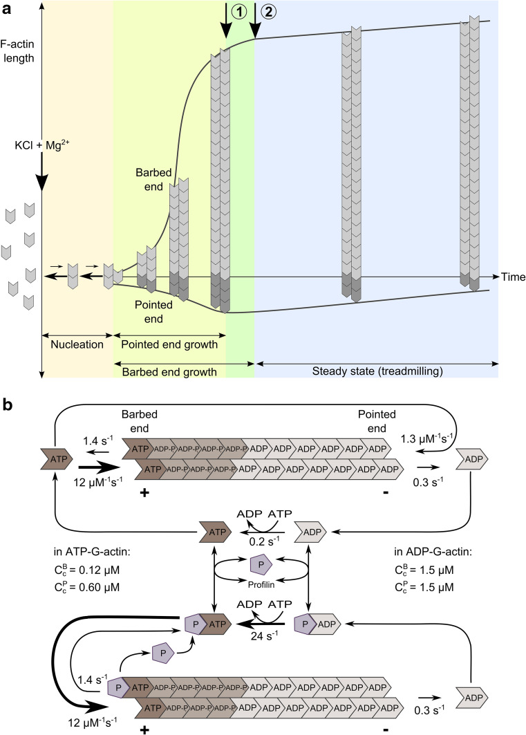
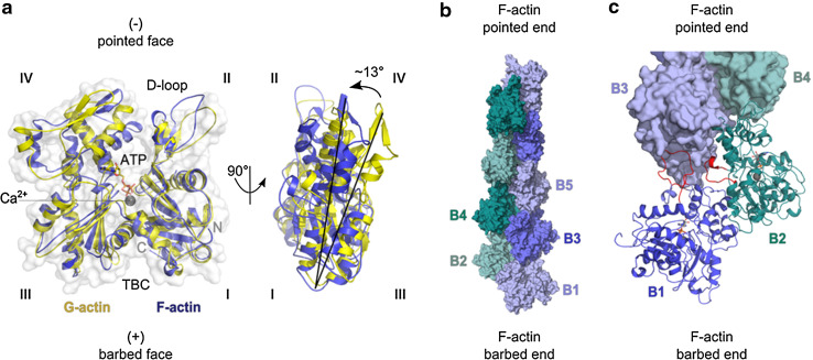
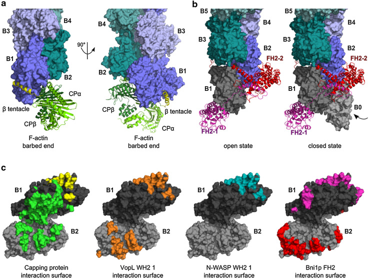
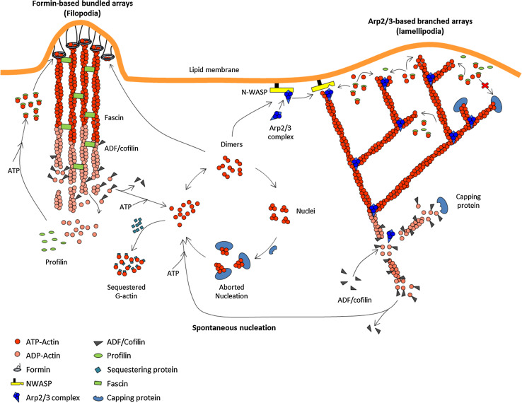
Actin cytoskeleton remodeling, which drives changes in cell shape and motility, is orchestrated by a coordinated control of polarized assembly of actin filaments. Signal responsive, membrane-bound protein machineries initiate and regulate polarized growth of actin filaments by mediating transient links with their barbed ends, which elongate from polymerizable actin monomers. The barbed end of an actin filament thus stands out as a hotspot of regulation of filament assembly. It is the target of both soluble and membrane-bound agonists as well as antagonists of filament assembly. Here, we review the molecular mechanisms by which various regulators of actin dynamics bind, synergize or compete at filament barbed ends. Two proteins can compete for the barbed end via a mutually exclusive binding scheme. Alternatively, two regulators acting individually at barbed ends may be bound together transiently to terminal actin subunits at barbed ends, leading to the displacement of one by the other. The kinetics of these reactions is a key in understanding how filament length and membrane-filament linkage are controlled. It is also essential for understanding how force is produced to shape membranes by mechano-sensitive, processive barbed end tracking machineries like formins and by WASP-Arp2/3 branched filament arrays. A combination of biochemical and biophysical approaches, including bulk solution assembly measurements using pyrenyl-actin fluorescence, single filament dynamics, single molecule fluorescence imaging and reconstituted self-organized filament assemblies, have provided mechanistic insight into the role of actin polymerization in motile processes.


Figures

Fig. 1
Fig. 1
Diagram of self-assembly of an “average” actin filament in vitro. a Spontaneous assembly of ATP-actin in vitro is initiated by a sudden increase in ionic strength at time zero, in a solution of actin monomers (G-actin). Nucleation is followed by endwise association of G-actin molecules to nuclei, faster at the barbed end than at the pointed end. As F-actin is assembled, the concentration of G-actin monomers decreases in solution. Pointed end growth rate reaches zero (arrow 1) when the concentration of G-actin reaches the critical concentration for pointed end assembly (0.6 µM). Barbed end growth goes on and G-actin concentration declines, while pointed ends start to disassemble. When the steady-state concentration of G-actin (0.1 µM) is reached (arrow 2), equal net rates of barbed end assembly and pointed end disassembly (treadmilling) maintain a constant amount of F-actin in solution, schematized here by a constant length of the “average” filament. b Nucleotide hydrolysis associated with the treadmilling cycle of the actin filament at steady-state in ATP, with and without profilin. The barbed end terminal subunits are enriched in ATP/ADP-Pi, while ADP is bound to pointed end terminal subunits. Profilin–ATP-actin participates in barbed end assembly, but not in pointed end assembly, hence it enhances processivity of treadmilling. In the cellular medium, treadmilling is regulated to generate variable rates of barbed end assembly. Regulation is performed either by increasing the rate limiting step of the treadmilling cycle which is pointed end depolymerization (using ADF/cofilin), or by regulating the dynamics at barbed ends (capping, tracking, destabilizing)
Fig. 2
Fig. 2
Structures of G-actin and F-actin filament barbed ends. a The transition of G- to F-actin. The structures of actin in the globular G-actin (yellow) or in the filamentous F-actin (blue) state are superimposed. They originate from G-actin in complex with DNAse I with bound Ca2+-ion and ATP (1ATN; [122]) and F-actin (2ZWH; [123]). The DNAse I-binding loop (D-loop) and subdomains IIV are labeled. The target binding cleft (TBC) at the barbed face of actin is located between subdomains I and III. Actin protomers are flattened in F-actin by a 13° twist of the outer subdomains (I and II) to the inner ones (III and IV). b Surface representation of the double-helical structure of a 167° twisted F-actin nonamer (4A7N; [124]). The fast growing barbed end and slow growing pointed end are indicated. c Important longitudinal contacts between terminal F-actin subunits at the filament barbed end. The two terminal actin protomers B1 and B2 are depicted as cartoon. Loops involved in intermolecular binding are highlighted (red). The D-loop of actin B1 (aa 31–51) including its adjacent C-terminal region (aa 61–65) binds into the TBC of protomer B3. It also contributes to the transverse interaction between loop aa 265–271 of B2 with B3. The longitudinal contact of loop aa 243–245 of B1 with B3 is not visible in this representation [9, 123]
Fig. 3
Fig. 3
Structure of actin regulators bound to the barbed end. a Structure of CP bound to the barbed end. The α/β heterodimeric capping protein (CP) is illustrated in ribbon diagrams (CPα: light green; CPβ: green; [43]; coordinates kindly provided by Y. Maeda). CPαβ forms strong electrostatic interactions at the interface of B1 and B2, while CPβ binds with its amphipathic β-tentacle (βT, yellow) to the hydrophobic TBC of B1. b Structure of a dimeric formin homology 2 (FH2) domain at the barbed end. The crystal structure of the FH2 domain of yeast formin Bni1p was crystallized encircling a flattened, 180° twisted pseudo filament (1Y64; [54]). The Bni1-FH2/G-actin structure was superimposed on actin B2 of the 167° twisted F-actin barbed end (4A7N, shown in greenblue). The 180° twisted protomers B1 and B0 are depicted in grey. The amphipathic α-helix of the knob region of each FH2 hemidimer (chains FH2-1, FH2-2; magenta, red) binds to the TBC of B1 and B2, respectively. c Interaction surface of actin regulators at the barbed end. Highlighted residues of B1 (dark grey) and B2 (grey) are involved in binding to the various regulators. Many barbed end binding proteins associate with an α-helix (e.g. β-tentacle of CP, yellow surface) to the TBC of actin and additionally with other surface areas specific for each interaction. Surface coloring: residues of the barbed end involved in binding to CP (CPαβ green, β-tentacle yellow), VopL-WH2 1 (3M1F, orange, aa 130–151; [125]), N-WASP-WH2 1 (3M3N, blue, aa 397–418; [125]), or Bni1p-FH2 domain (1Y64, magenta, red; [54]). Since VopL dimerizes, the interacting residues of the first, N-terminal WH2 domain of each VopL chain were highlighted on B1 and B2, respectively. The diagram suggests that two barbed end binding regulators can bind together to B1 and B2 pending some loss of binding strength, and use this transient ternary complex to displace each other. Examples include uncapping of CP by VopF [21] and by formin (Shekhar et al., submitted)
Fig. 4
Fig. 4
Sketch of the regulation of filament assembly in motile processes. Regulated treadmilling drives both site-directed barbed end nucleation and polarized assembly. For simplicity, only the protein machineries responsible for filament branching (WASP family proteins) and for processive individual filament assemblies (formins) are drawn. Filament tracking by Ena/VASP and other WH2 domain proteins are conceptually similar, and not shown for simplicity. In the generalized treadmilling cycle, polymerizable ATP-bound actin monomers are produced by depolymerization of ADP-actin from filament ADP-bound pointed ends, facilitated by ADF/cofilin. Note that an excess of ADF will block monomers in the ADF–ADP-bound non-motile state (no treadmilling), because nucleotide exchange is inhibited by ADF. Thus, the effect of ADF on motility presents a bell shape dependence on concentration. Spontaneous nucleation by ATP-actin is aborted in cytoplasm by capping protein, and locally facilitated by nucleators. Formin-induced nucleation requires actin dimers. The sketch implicitly assumes that an actin dimer/trimer prenucleus can as well undergo branching with WASP and Arp2/3 complex. Capping protein arrests filament growth in dendritic filament arrays. A balanced number of filament barbed ends is maintained via the equal frequency of “birth” by branching and “death” by capping. Capping protein is also required for regulating the length of formin-induced filament in filopodia

References

    1. Pollard TD, Borisy GG. Cellular motility driven by assembly and disassembly of actin filaments. Cell. 2003;112(4):453–465. - PubMed
    1. Welch MD, Way M. Arp2/3-mediated actin-based motility: a tail of pathogen abuse. Cell Host Microbe. 2013;14(3):242–255. - PMC - PubMed
    1. Iwasa JH, Mullins RD. Spatial and temporal relationships between actin-filament nucleation, capping, and disassembly. Curr Biol. 2007;17(5):395–406. - PMC - PubMed
    1. Lai FP, Szczodrak M, Block J, Faix J, Breitsprecher D, Mannherz HG, Stradal TE, Dunn GA, Small JV, Rottner K. Arp2/3 complex interactions and actin network turnover in lamellipodia. EMBO J. 2008;27(7):982–992. - PMC - PubMed
    1. Bugyi B, Carlier MF. Control of actin filament treadmilling in cell motility. Annu Rev Biophys. 2010;39:449–470. - PubMed

Publication types

MeSH terms

Substances

LinkOut - more resources