

Review
doi: 10.1007/s00018-015-1917-z.
Epub 2015 May 7.
Atg1 family kinases in autophagy initiation
Affiliations
- PMID: 25948417
- PMCID: PMC4506457
- DOI: 10.1007/s00018-015-1917-z
Item in Clipboard
Review
Atg1 family kinases in autophagy initiation
Cell Mol Life Sci.
2015 Aug.
Abstract
Autophagosome formation, a landmark event in autophagy, is accomplished by the concerted actions of Atg proteins. Among all Atg proteins, Atg1 kinase in yeast and its counterpart in higher eukaryotes, ULK1 kinase, function as the most upstream factor in this process and mediate autophagy initiation. In this review, we summarize current knowledge of the structure, molecular function, and regulation of Atg1 family kinases in the initiation of autophagy.
Figures
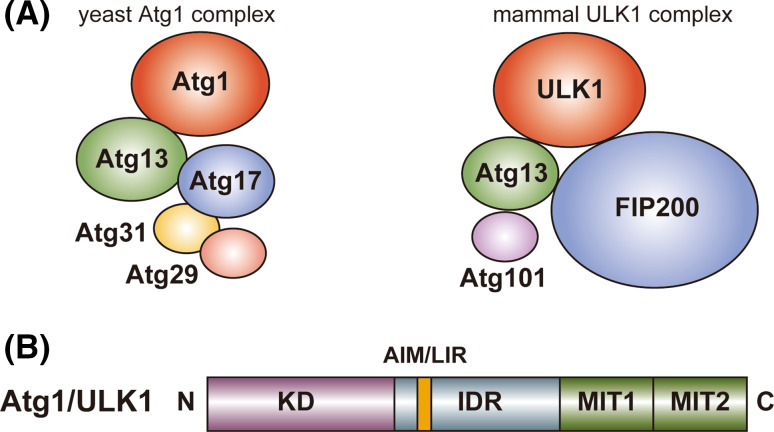
Schematic drawing of the domain organization and the binding partners of Atg1/ULK1. a Yeast (S. cerevisiae) Atg1 and mammalian ULK1 complexes. b Domain organization of Atg1/ULK1. N and C show amino- and carboxy-terminus, respectively
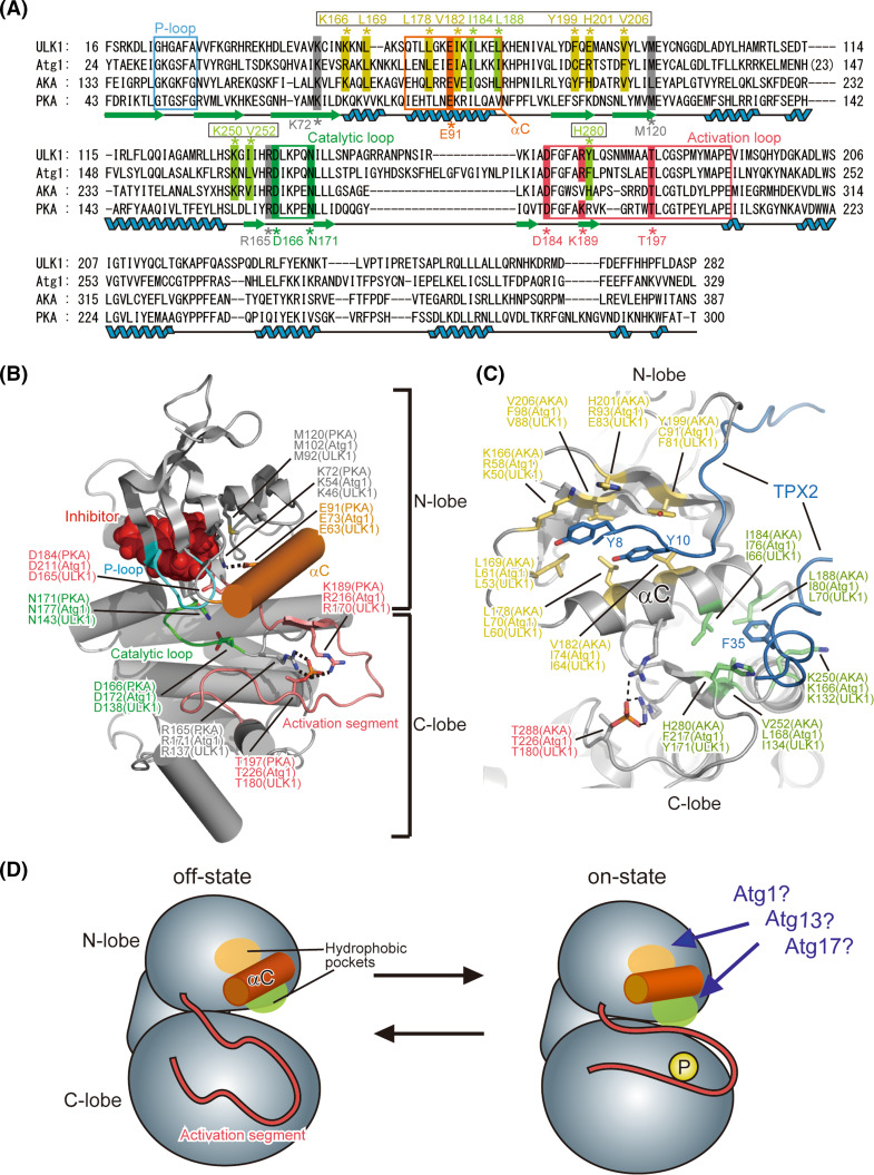
Proposed architecture of Atg1/ULK1 KD. a Sequence alignment among Atg1, ULK1, AKA and PKA. Secondary structure elements of PKA are denoted under the sequence. Unboxed residue numbers indicated below the sequence correspond to those of PKA, whereas boxed residue numbers indicated above the sequence correspond to those of AKA. b Crystal structure of the kinase domain of ULK1 (PDB ID 4WNO) [101]. The side chains of the catalytically important residues are shown with a stick model. Broken lines indicate possible salt bridges that are important for the on-state of the kinase. All structural models in this manuscript were prepared using the program PyMOL [102]. c Crystal structure of AKA in complex with TPX2 (PDB ID 1OL5). The side-chain of the residues that constitute the two hydrophobic pockets accommodating TPX2 is shown with a stick model. d Schematic drawing of the conformational switch of Atg1 KD between the off-state and on-state
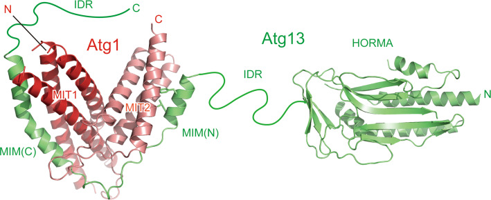
Architecture of Atg13 and tandem MIT domains of Atg1. Ribbon models are prepared using the crystal structures of Atg13 HORMA (PDB ID 4J2G) and two MIMs bound to the two MIT domains of Atg1 (PDB ID 4P1N). The side-chain of the two hydrophobic residues in MIM(N) that are important for Atg1 binding is shown with a stick model
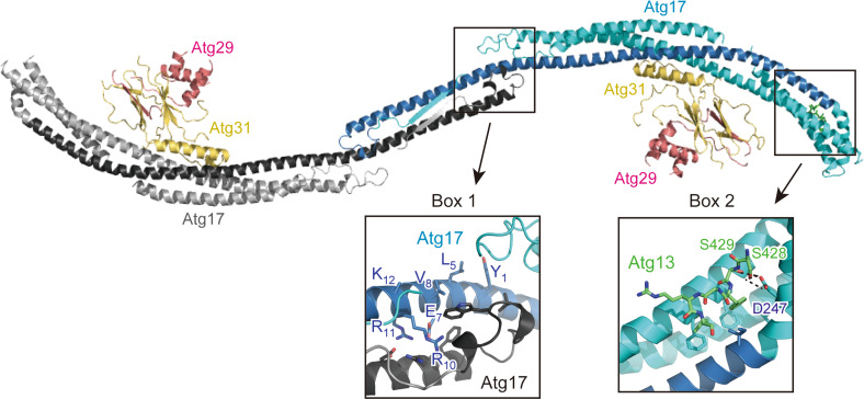
Architecture of the Atg17–Atg29–Atg31 complex and its interaction with Atg1317BR. Ribbon models were prepared using the crystal structure of the Atg1317BR–Atg17–Atg29–Atg31 complex (PDB ID 4P1W). Two protomers of Atg17 are colored cyan and gray, in which the regions that show weak homology with Atg11 and FIP200 are colored blue and black, respectively. Boxes 1 and 2 indicate a close-up view of the dimer interphase and the 17BR-binding site, respectively
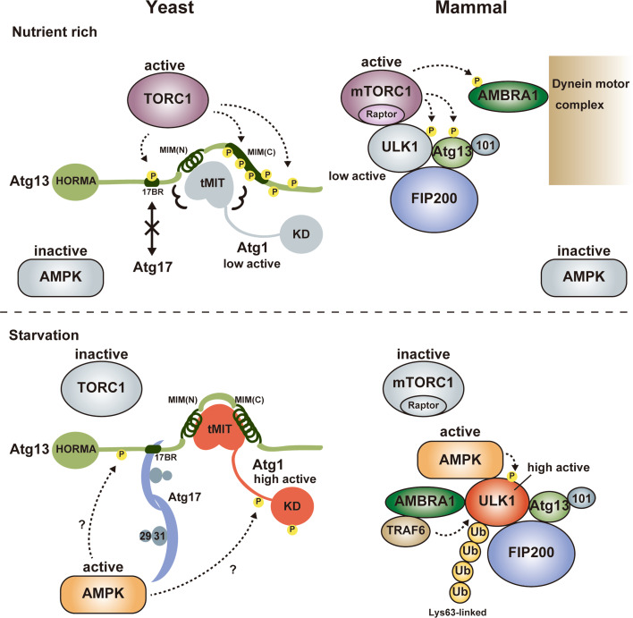
Summary of Atg1/ULK1 regulation. Under nutrient-rich conditions, TORC1 is active and directly phosphorylates Atg13 at 17BR and MIM(C), which impairs the formation of the Atg1 complex in yeast. In mammals, mTORC1 binds to ULK1 and phosphorylates ULK1, Atg13 and AMBRA1, which keeps the ULK1 complex as an inactive state although it does not impair the formation of the ULK1 complex. Upon starvation, TORC1 is inactive and Atg13 is dephosphorylated, which leads to the formation of the Atg1 complex in yeast. Activated AMPK also positively regulates the Atg1 complex possibly via phosphorylation of Atg1 and/or Atg13. In mammals, mTORC1 dissociates from the ULK1 complex and ULK1 is activated, which is also positively regulated by Lys63-linked ubiquitylation by the AMBRA1-TRAF6 complex and phosphorylation by activated AMPK. Circled P indicates phosphorylation sites
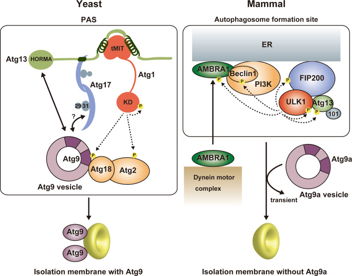
Summary of autophagy initiation mediated by Atg1/ULK1. In yeast, Atg9 vesicle is recruited to the PAS via the interaction with Atg13 HORMA and the Atg17–Atg9 interaction might also be responsible for that. Activated Atg1 phosphorylates Atg9, which promotes the recruitment of downstream factors such as Atg18 to the PAS. Atg9 is integrated into the isolation membrane. In mammals, activated ULK1 phosphorylates many factors, among which phosphorylation of Beclin 1 and AMBRA1 is responsible for the targeting of the ULK1 complex and the PI3 K complex to the autophagosome formation site at ER. In contrast to yeast Atg9, mammalian Atg9a targets to the autophagosome formation site independently of other Atg factors and transiently, and is not integrated into the isolation membrane
References
Publication types
MeSH terms
Substances
LinkOut - more resources
Full Text Sources
Other Literature Sources
Molecular Biology Databases
