. 2015 May 7:13:147.
doi: 10.1186/s12967-015-0510-4.

Fibroblast growth factor-2 regulates human cardiac myofibroblast-mediated extracellular matrix remodeling

Affiliations

Fibroblast growth factor-2 regulates human cardiac myofibroblast-mediated extracellular matrix remodeling

Daniyil A Svystonyuk et al. J Transl Med. .

Abstract

Background: Tissue fibrosis and chamber remodeling is a hallmark of the failing heart and the final common pathway for heart failure of diverse etiologies. Sustained elevation of pro-fibrotic cytokine transforming growth factor-beta1 (TGFβ1) induces cardiac myofibroblast-mediated fibrosis and progressive structural tissue remodeling.

Objectives: We examined the effects of low molecular weight fibroblast growth factor (LMW-FGF-2) on human cardiac myofibroblast-mediated extracellular matrix (ECM) dysregulation and remodeling.

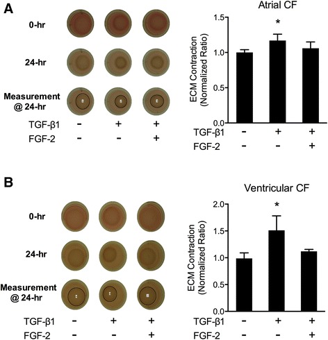
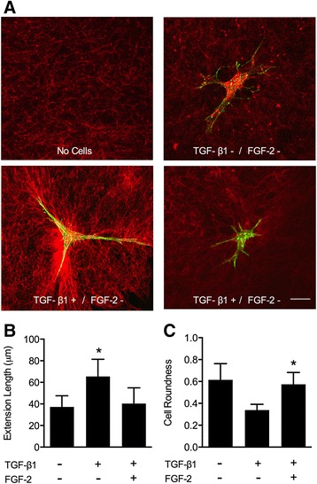
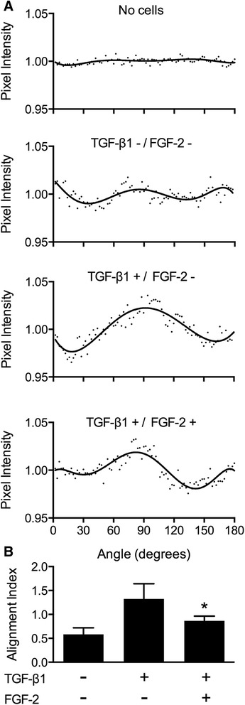
Methods: Human cardiac biopsies were obtained during open-heart surgery and myofibroblasts were isolated, passaged, and seeded within type I collagen matrices. To induce myofibroblast activation and ECM remodeling, myofibroblast-seeded collagen gels were exposed to TGFβ1. The extent of ECM contraction, myofibroblast activation, ECM dysregulation, and cell apoptosis was determined in the presence of LMW-FGF-2 and compared to its absence. Using a novel floating nylon-grid supported thin collagen gel culture platform system, myofibroblast activation and local ECM remodeling around isolated single cells was imaged using confocal microscopy and quantified by image analysis.

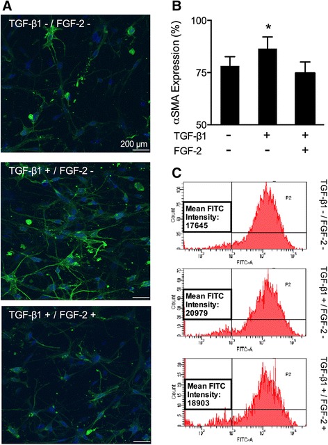
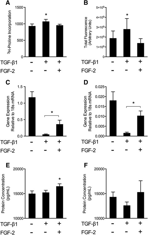
Results: TGFβ1 induced significant myofibroblast activation and ECM dysregulation as evidenced by collagen gel contraction, structural ECM remodeling, collagen synthesis, ECM degradation, and altered TIMP expression. LMW-FGF-2 significantly attenuated TGFβ1 induced myofibroblast-mediated ECM remodeling. These observations were similar using either ventricular or atrial-derived cardiac myofibroblasts. In addition, for the first time using individual cells, LMW-FGF-2 was observed to attenuate cardiac myofibroblast activation and prevent local cell-mediated ECM perturbations.

Conclusions: LMW-FGF-2 attenuates human cardiac myofibroblast-mediated ECM remodeling and may prevent progressive maladaptive chamber remodeling and tissue fibrosis for patients with diverse structural heart diseases.


Figures

Figure 1
Figure 1
Extracellular matrix remodeling by human cardiac myofibroblasts. TGFβ1 stimulates both atrial (A) and ventricular (B) myofibroblast-mediated collagen gel contraction as compared to controls. This effect was inhibited in the presence of LMW-FGF-2. Data presented were obtained from multiple individual experiments (4 experiments, total N = 14 per group for A; 3 experiments, total N = 8 per group for B) and all values were normalized to the corresponding control groups. Bars represent mean ± SD; *p < 0.01, **p < 0.001; abbreviation: CF – cardiac fibroblasts.
Figure 2
Figure 2
Effects of LMW-FGF-2 on myofibroblast activation. (A) Representative confocal microscopic images of cardiac myofibroblasts embedded in a collagen gel, following treatments (control, TGFβ1 and LMW-FGF-2 + TGFβ1). Cells were stained for α-SMA (green) and nuclei (DAPI; blue). (B) TGFβ1 increased α-SMA expression while LMW-FGF-2 diminished α-SMA expression. Bars represent mean ± SD; *p < 0.01. (C) α-SMA was labeled with FITC-conjugated antibody and the intensity of a-SMA expression was measured using flow cytometry. TGFβ1 increased the cellular expression of α-SMA and this effect was abolished by LMW-FGF-2.
Figure 3
Figure 3
ECM regulation by collagen synthesis, protease activity, and TIMP inhibition. (A) Collagen synthesis was assessed by measurement of 3H-proline incorporation. TGFβ1 increased 3H-proline incorporation. This effect was abolished by addition of LMW-FGF-2. (B) ECM degradation was assessed by in situ zymography using embedded DQ™ Gelatin-FITC to measure matrix proteolysis. Total protease activity was quantified as total fluorescent signal per image volume. TGFβ1 increased total protease activity while LMW-FGF-2 attenuated this response. (C and D) Expression of TIMP-1 and TIMP-2, respectively, at the mRNA levels are presented. TGFβ1 significantly reduced both TIMP-1 and TIMP-2 expression while LMW-FGF-2 partially restored expression. (E) At the protein level, TGFβ1 did not alter TIMP-1, however the presence of LMW-FGF-2 resulted in increased TIMP-1 protein. (F) Though the results for TIMP-2 were not statistically significant, a trend toward decreased TIMP-2 protein was noted in the TGFβ1 group and restored in the presence of LMW-FGF-2. *, p < 0.05.
Figure 4
Figure 4
Myofibroblast morphology: extension length and roundness. (A) Representative confocal images of single isolated human cardiac myofibroblasts embedded into a thin collagen nylon-based gel. Following treatments (control, TGFβ1, TGFβ1 + LMW-FGF-2), cells were stained for F-actin (phalloidin; green) while adjacent collagen matrix was visualized using confocal reflectance (red). (B) Cell extension length was measured from the cell centroid to the tip of a given cell extension. TGFβ1 increased mean cell extension length while LMW-FGF-2 restored levels close to baseline. Bars represent mean ± SD; *p < 0.05. (C) Roundness, a dimensionless shape factor, was quantified for each treatment group. Values closer to 1.0 represent round phenotypes while decreasing values <1.0 are representative of a star-shaped phenotype (activated). TGFβ1 treatment favored a more stellate-cell shape while LMW-FGF-2 showed morphology consistent with non-activated cells. Bars represent mean ± SD; *p < 0.05. Scale bar = 20 μm.
Figure 5
Figure 5
Extracellular matrix remodeling: collagen fiber alignment. Collagen fiber alignment was assessed parallel to cell extensions as a measure of cell-mediated ECM remodeling. (A) Normalized pixel intensity curves generated from FFT analysis for each treatment group relative to a no-cell control. Randomly oriented collagen fibers result in constant pixel intensity at any given angle. Alignment of fibers parallel to cell extensions result in increased intensity at certain angles. (B) Alignment index is calculated by taking the area under the curve ± 10 degrees from the maximum intensity value. TGFβ1 showed the greatest collagen fiber alignment while LMW-FGF-2 attenuated this response. Bars represent mean ± SD; *p < 0.05.

Similar articles

Cited by

References

    1. Fedak PW, Verma S, Weisel RD, Li RK. Cardiac remodeling and failure: from molecules to man (Part I) Cardiovasc Pathol. 2005;14(1):1–11. doi: 10.1016/j.carpath.2004.12.002. - DOI - PubMed
    1. Spinale FG. Matrix metalloproteinases: regulation and dysregulation in the failing heart. CircRes. 2002;90(5):520–30. - PubMed
    1. Biernacka A, Dobaczewski M, Frangogiannis NG. TGF-β signaling in fibrosis. Growth factors. 2011;29:196–202. doi: 10.3109/08977194.2011.595714. - DOI - PMC - PubMed
    1. Dobaczewski M, Chen W, Frangogiannis NG. Transforming growth factor (TGF)-β signaling in cardiac remodeling. J Mol Cell Cardiol. 2011;51(4):600–6. doi: 10.1016/j.yjmcc.2010.10.033. - DOI - PMC - PubMed
    1. Leask A. Potential therapeutic targets for cardiac fibrosis: TGFbeta, angiotensin, endothelin, CCN2, and PDGF, partners in fibroblast activation. Circ Res. 2010;106:1675–80. doi: 10.1161/CIRCRESAHA.110.217737. - DOI - PubMed

Publication types

Substances