. 2015 May 6;4(5):e001920.
doi: 10.1161/JAHA.115.001920.

Tumor Necrosis Factor-α Underlies Loss of Cortical Dendritic Spine Density in a Mouse Model of Congestive Heart Failure

Affiliations

Tumor Necrosis Factor-α Underlies Loss of Cortical Dendritic Spine Density in a Mouse Model of Congestive Heart Failure

Anja Meissner et al. J Am Heart Assoc. .

Abstract

Background: Heart failure (HF) is a progressive disorder characterized by reduced cardiac output and increased peripheral resistance, ultimately leading to tissue perfusion deficits and devastating consequences for several organs including the brain. We previously described a tumor necrosis factor-α (TNF-α)-dependent enhancement of posterior cerebral artery tone and concomitant reduced cerebral blood flow in a mouse model of early HF in which blood pressure remains minimally affected. HF is often associated with cognitive impairments such as memory deficits, even before any overt changes in brain structure and function occur. The pathophysiology underlying the development of cognitive impairments in HF is unknown, and appropriate treatment strategies are lacking.

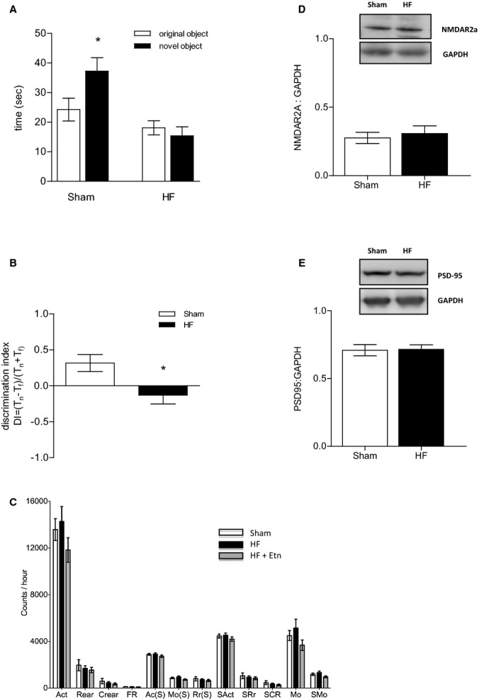
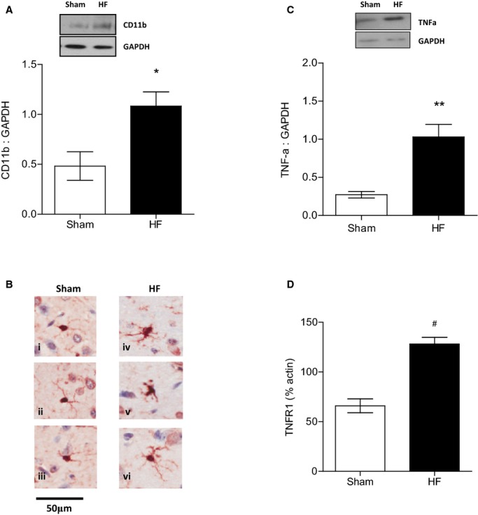
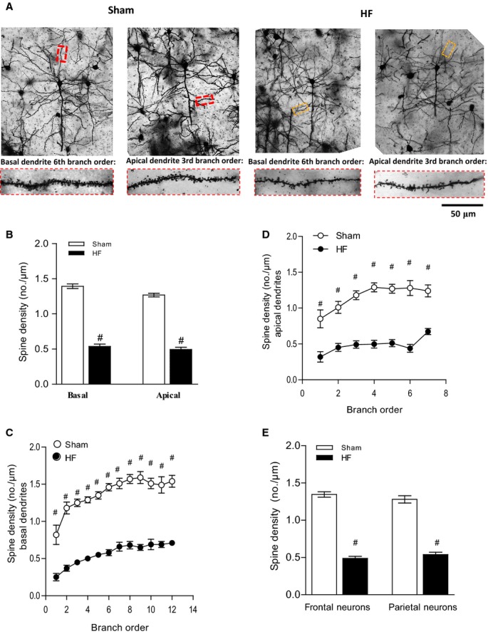
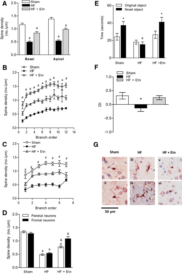
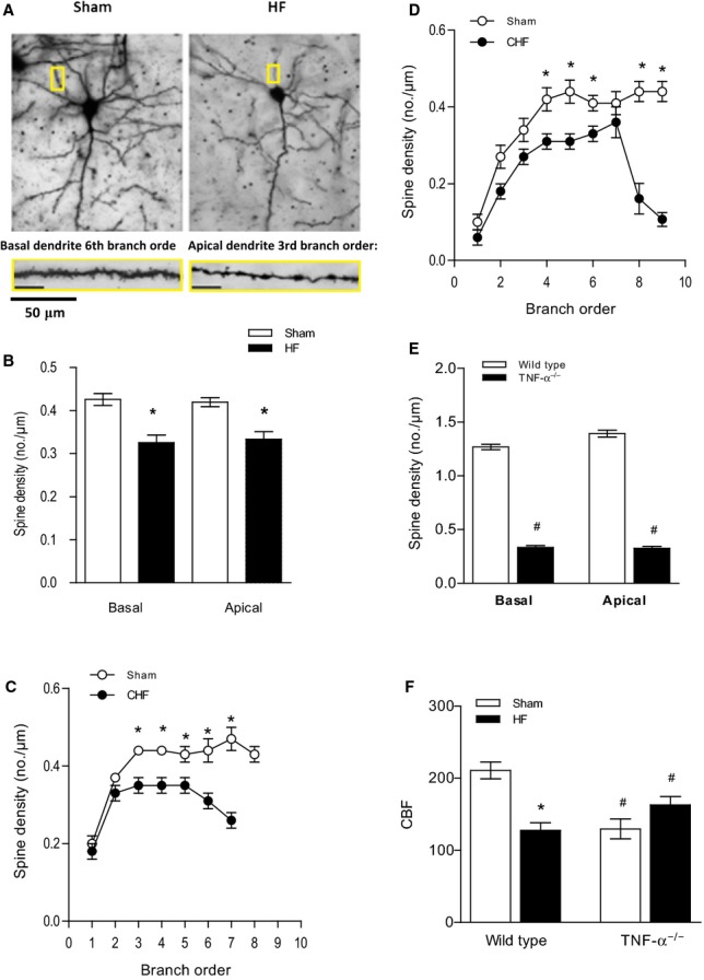
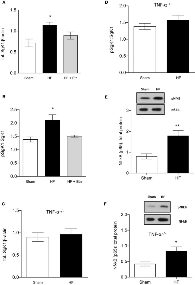
Methods and results: We used a well-established mouse model in which HF was induced by experimental myocardial infarction produced by permanent surgical ligation of the left anterior descending coronary artery (infarct size ≈25% of the left ventricular wall). Ligated mice developed enlarged hearts, congested lungs, and reduced cardiac output and blood pressure, with elevated peripheral resistance within 6 to 8 weeks after ligation. In this study, we demonstrated the significance of the proinflammatory cytokine TNF-α during HF-mediated neuroinflammation and associated impaired hippocampus-independent nonspatial episodic memory function. Augmented cerebral TNF-α expression and microglial activation in HF mice, indicative of brain inflammation, were accompanied by morphological changes and significant reduction of cortical dendritic spines (61.39±8.61% for basal and 61.04±9.18% for apical spines [P<0.001]). The significance of TNF-α signaling during the observed HF-mediated neurodegenerative processes is supported by evidence showing that sequestration or genetic deletion of TNF-α ameliorates the observed reduction of cortical dendritic spines (33.51±7.63% for basal and 30.13±6.98% for apical spines in wild-type mice treated with etanercept; 17.09±6.81% for basal and 17.21±7.29% for apical spines in TNF-α(-/-)). Moreover, our data suggest that alterations in cerebral serum and glucocorticoid-inducible kinase 1 (SgK1) expression and phosphorylation during HF may be TNF-α dependent and that an increase of SgK1 phosphorylation potentially plays a role in the HF-associated reduction of dendritic spine density.

Conclusions: Our findings demonstrate that TNF-α plays a pivotal role in HF-mediated neuroinflammation and associated alterations of cortical dendritic spine density and has the potential to reveal novel treatment strategies for HF-associated memory deficits.

Keywords: heart failure; inflammation; nervous system.


Figures

Figure 1
Figure 1
HF attenuates hippocampus-independent, short-term memory. A, HF attenuated the rodents’ short-term retention of object familiarity in a nonspatial novel object recognition task with a 5-minute retention interval. B, The calculated DI revealed a clear preference for the familiar rather than the novel object in HF mice. C, All aspects of locomotor activity were shown not to be significantly different between sham and HF mice. For all tasks: sham n=9, HF n=10,*P<0.05. D, Cerebral protein expression of NMDAR2A (n=7, P=0.6494) and (E) PSD-95 (n=6, P=0.9074) was not affected during HF. Ac(S) indicates active time; Act, activity; Crear, center rearing; DI, discrimination index; Etn, etanercept; FR, front to back count; HF, heart failure; Mo(S), mobile time; MO, mobile counts; NMDAR, N-methyl-D-aspartate receptor; PSD-95, postsynaptic density protein 95; Rear, rearing; Rr(S), rearing time; SAct, slow activity; SCR, slow center rearing; SMO, slow mobile counts; SRr, slow rearing.
Figure 2
Figure 2
Memory deficits of HF mice are accompanied by increased cerebral TNF-α levels and activated microglia. A, Increased expression of CD11b, the β-integrin marker of microglia, represents microglial activation during neurodegenerative inflammation during HF (n=5, P<0.05). B, Cerebral tissue sections of HF mice show evidence of microglial activation with microglia exhibiting visibly enlarged cytoplasm and reduced, thickened processes (iv through vi) compared with sham-operated animals (i through iii). C, TNF-α protein levels in the brain were significantly increased in mice 8 to 10 weeks after myocardial infarction compared with sham controls (n=5, P<0.01). D, TNF-α receptor type 1 mRNA was significantly upregulated during HF compared with sham controls (n=6, P<0.001). *P<0.05, **P<0.01 and #P<0.001. HF indicates heart failure; TNF-α, tumor necrosis factor-α.
Figure 3
Figure 3
HF induces morphological changes and a reduction in number of dendritic spines. The sample population was composed of pyramidal cells chosen rostrocaudally from motor, sensory, and visual cortical regions (4 to 6 cells per animal). A, Depicts representative Golgi-stained images of cortical neurons of sham and HF wild-type mice and illustrates the differences between the basal and apical neurons of sham mice, which had significant more dendritic branches and thicker branch diameters than those of HF mice. The magnification shows details of both basal and apical dendrite morphology. B, Spine density of both basal and apical dendrites was reduced in the frontal cortex in HF mice. Breakdown analysis (dendrograms) further revealed a significant loss of spines or spine density in both (C) basal and (D) apical dendrites of HF compared with sham. E, Spine density of frontal neurons and parietal neurons is significantly reduced during HF. For all tasks: sham n=18, HF n=20, #P<0.001. HF indicates heart failure.
Figure 4
Figure 4
TNF-α scavenger etanercept partially rescues HF-induced effects in the brain. A, Dendritic spine density was found to be reduced in the frontal cortex in HF mice treated with Etn compared with sham controls but significantly increased compared with untreated HF mice. Breakdown analysis (dendrograms) further revealed significant differences of spines or spine density in both (B) basal and (C) apical dendrites of Etn-treated HF compared with HF mice. D, Spine density of frontal and parietal neurons is significantly reduced during HF. Etn fully prevented HF-mediated reduction in the frontal neurons but only partially prevented it in the parietal neurons. For all tasks: sham n=18, HFn=20, HFplus Etn n=13. #P<0.001 between control and HF, &P<0.001 between HF and HF plus Etn. E, In a nonspatial novel object recognition task with a 5-minute retention period, HF animals treated with Etn spent more time exploring the novel object and (F) showed a clear preference for novelty compared with untreated mice. For all tasks: sham n=9, HF n=10, HFplus Etn n=10. *P<0.05 and #P<0.001. G, Cerebral tissue sections of HF mice treated with Etn show, similar to control mice (i and ii), no evidence of microglial activation (v and vi) compared with untreated HF animals, which exhibit microglia with visibly enlarged cytoplasm and reduced, thickened processes (iii and iv) (n=3). DI indicates discrimination index; Etn, etanercept; HF, heart failure; TNF-α, tumor necrosis factor-α.
Figure 5
Figure 5
HF-induced effects in the brain are ameliorated in TNF-α−/− mice. A, Representative images of dendritic spines of pyramidal cells from TNF-α−/− mice. The magnification shows details of both basal and apical dendrite morphology (5 to 7 cells per animal). B, Total spine density of basal and apical dendrites was significantly reduced in HF mice (4 to 6 cells per animal). Breakdown analysis of spine density found that significant reductions occurred across several branch orders of (C) basal dendrites and (D) apical dendrites between sham and HF mice. D, The average spine density of TNF-α−/− sham mice was significantly lower than that of normal wild-type animals in both basal and apical dendrites. For all tasks: sham n=24, HF n=24. *P<0.05, #P<0.001. E, HF had no effect on cerebral blood flow in TNF-α−/− mice (n=5, P=0.9069) compared with TNF-α−/− sham-operated mice. F, CBF, shown as mL/(100 g×minute), was significantly reduced compared with wild-type control animals (n=5, P<0.001). *P<0.05 within a group, #P<0.05 between wild-type and TNF-α−/− mice. CBF indicates cerebral blood flow; CHF, congestive heart failure; HF, heart failure; TNF-α, tumor necrosis factor-α.
Figure 6
Figure 6
SgK1 mRNA levels, protein expression, and phosphorylation status are altered during HF. A, During HF, SgK1 mRNA levels were upregulated compared with sham mice (n=6, P<0.05). Etn-treated HF mice showed SgK1 mRNA expression similar to that of untreated HF mice (n=6, P=0.2). B, HF induced an increase in SgK1 protein phosphorylation, which is significantly reduced after treating HF mice with Etn (n=5, P<0.05). HF had no effect on (C) SgK1 mRNA levels in TNF-α−/− mice (n=5, P=0.74) or (D) on SgK1 protein phosphorylation (n=5, P=0.38). Cerebral NF-kB (p65) was found to be activated during HF in (E) wild-type mice (n=5, versus <0.01) and (F) TNF-α deficient mice (n=5, P<0.01). *P<0.05 and **P<0.01. Etn indicates etanercept; HF, heart failure; NF-kB, nuclear factor-κB; p, phosphorylated; SgK1, serum and glucocorticoid-inducible kinase 1; TNF-α, tumor necrosis factor-α.

References

    1. Malik AS, Giamouzis G, Georgiopoulou VV, Fike LV, Kalogeropoulos AP, Norton CR, Sorescu D, Azim S, Laskar SR, Smith AL, Dunbar SB, Butler J. Patient perception versus medical record entry of health-related conditions among patients with heart failure. Am J Cardiol. 2011;107:569–572. - PMC - PubMed
    1. Havranek EP, Dwinnell B, Smith KS. Multiorgan failure after cardiac arrest in a 20-year-old man. Circulation. 1995;92:3139–3143. - PubMed
    1. Bennett SJ, Oldridge NB, Eckert GJ, Embree JL, Browning S, Hou N, Chui M, Deer M, Murray MD. Comparison of quality of life measures in heart failure. Nurs Res. 2003;52:207–216. - PubMed
    1. Vogels RL, Oosterman JM, van Harten B, Gouw AA, Schroeder-Tanka JM, Scheltens P, van der Flier WM, Weinstein HC. Neuroimaging and correlates of cognitive function among patients with heart failure. Dement Geriatr Cogn Disord. 2007;24:418–423. - PubMed
    1. Marshall RS, Lazar RM. Pumps, aqueducts, and drought management: vascular physiology in vascular cognitive impairment. Stroke. 2011;42:221–226. - PMC - PubMed

Publication types

MeSH terms

Substances