. 2015 Aug 1;24(15):4284-95.
doi: 10.1093/hmg/ddv160. Epub 2015 May 6.

Disruption of the lamin A and matrin-3 interaction by myopathic LMNA mutations

Affiliations

Disruption of the lamin A and matrin-3 interaction by myopathic LMNA mutations

Frederic F Depreux et al. Hum Mol Genet. .

Abstract

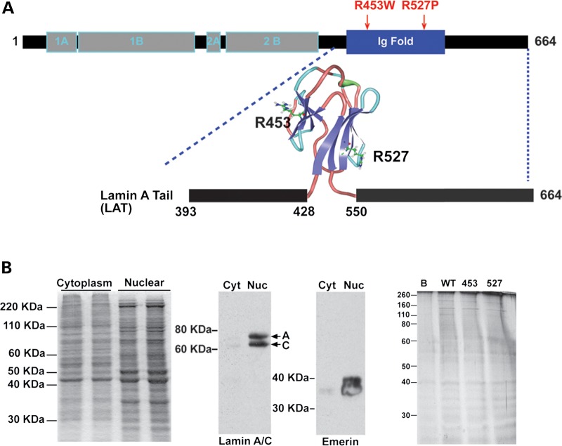
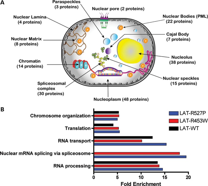
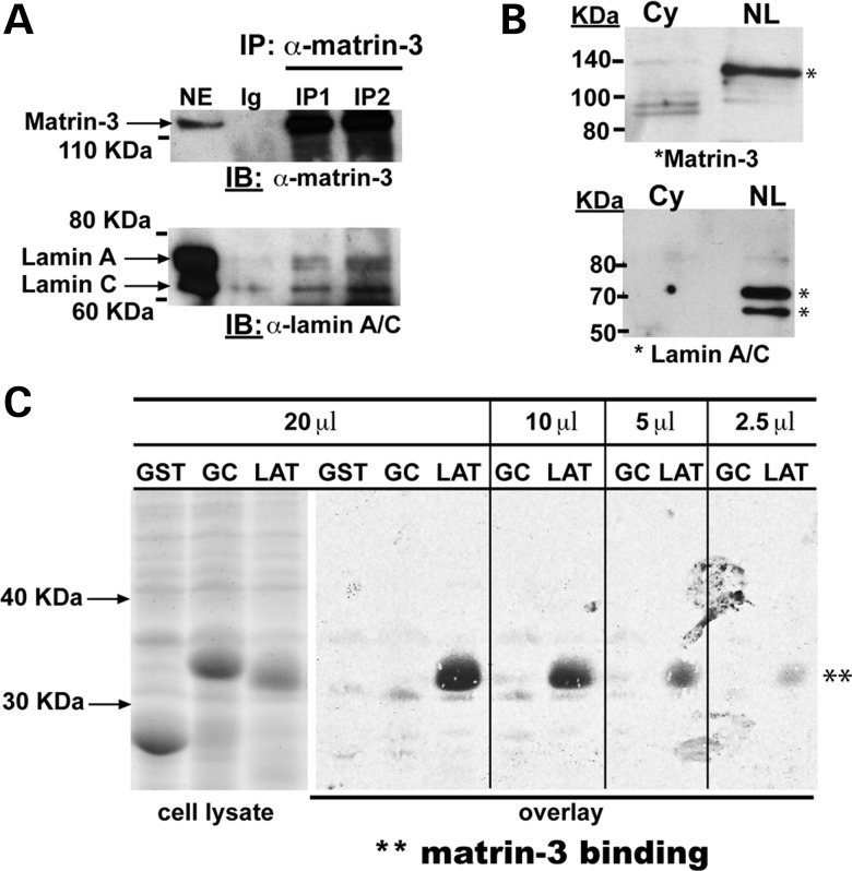
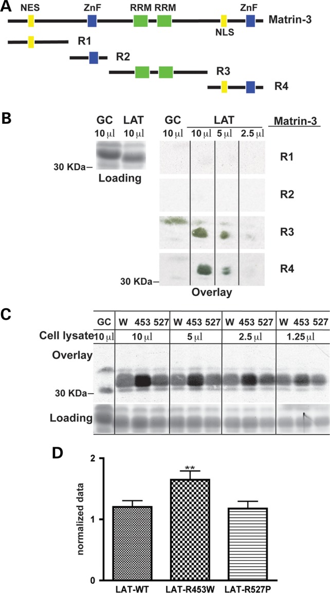
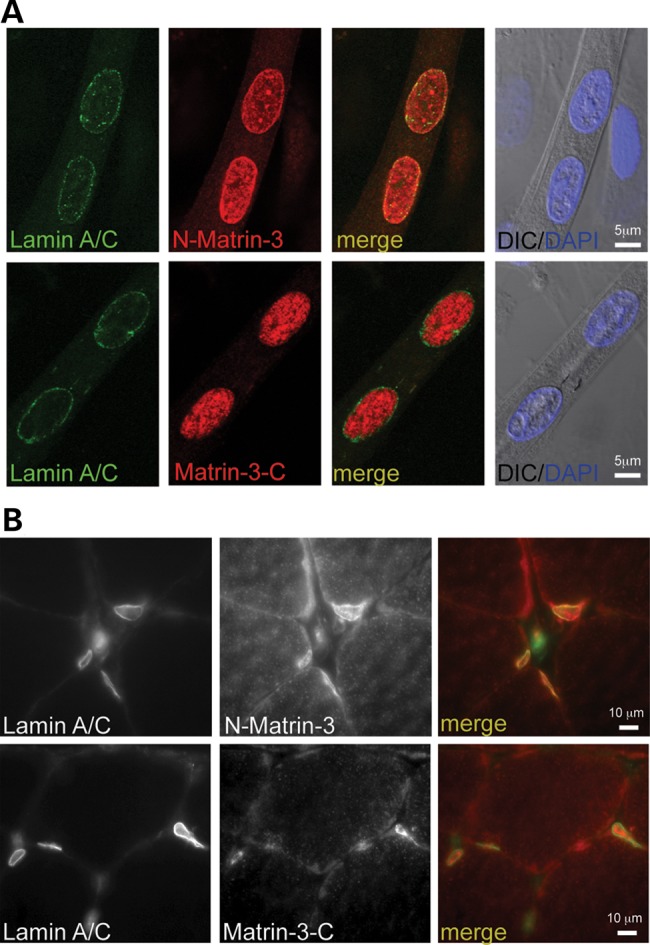
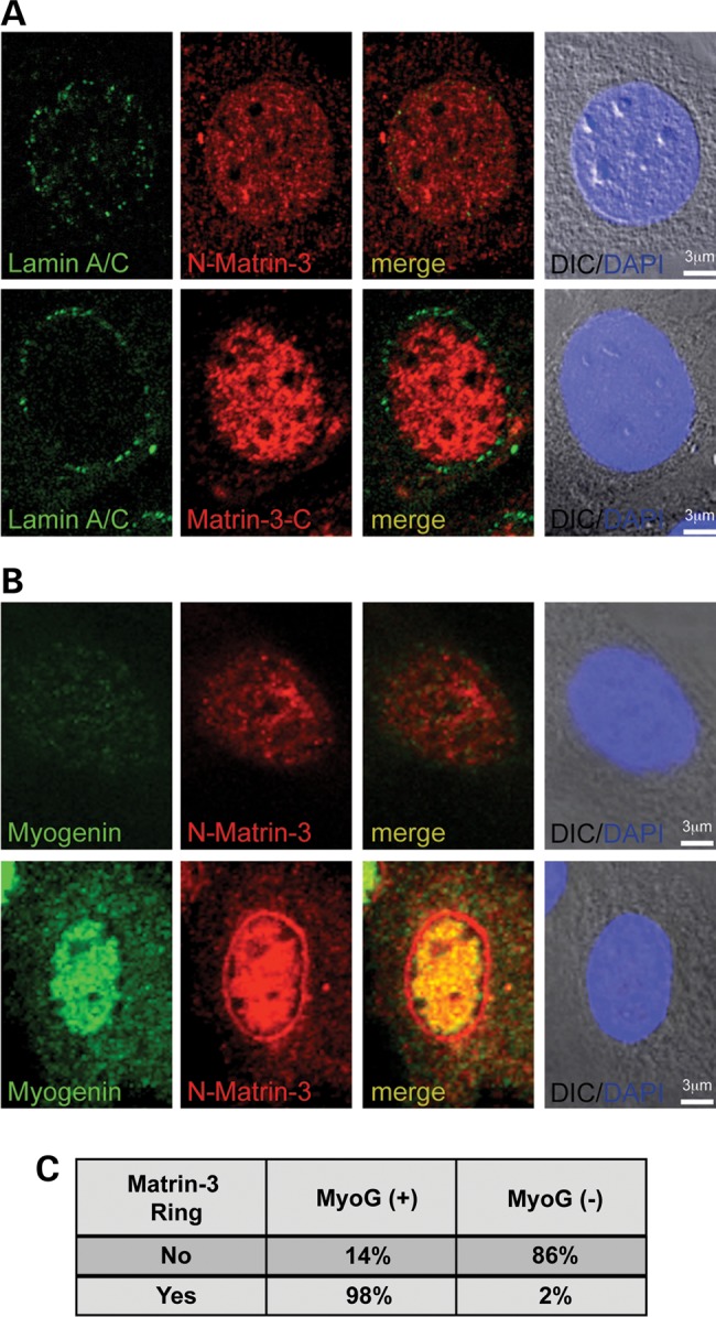
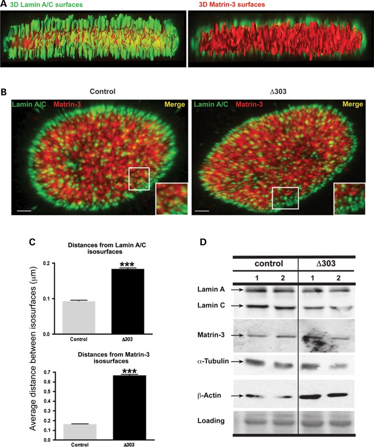
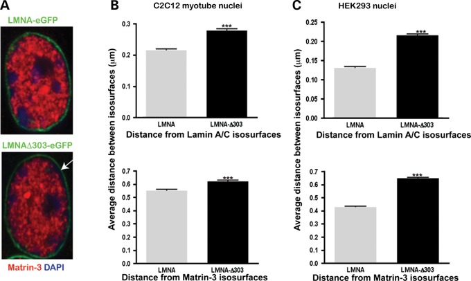
The nuclear face of the nuclear membrane is enriched with the intermediate filament protein lamin A. Mutations in LMNA, the gene encoding lamin A, lead to a diverse set of inherited conditions including myopathies that affect both the heart and skeletal muscle. To gain insight about lamin A protein interactions, binding proteins associated with the tail of lamin A were characterized. Of 130 nuclear proteins found associated with the lamin A tail, 17 (13%) were previously described lamin A binding partners. One protein not previously linked to lamin A, matrin-3, was selected for further study, because like LMNA mutations, matrin-3 has also been implicated in inherited myopathy. Matrin-3 binds RNA and DNA and is a nucleoplasmic protein originally identified from the insoluble nuclear fraction, referred to as the nuclear matrix. Anti-matrin-3 antibodies were found to co-immunoprecipitate lamin A, and the lamin-A binding domain was mapped to the carboxy-terminal half of matrin-3. Three-dimensional mapping of the lamin A-matrin-3 interface showed that the LMNA truncating mutation Δ303, which lacks the matrin-3 binding domain, was associated with an increased distance between lamin A and matrin-3. LMNA mutant cells are known to have altered biophysical properties and the matrin-3-lamin A interface is positioned to contribute to these defects.


Figures

Figure 1.
Figure 1.
Expression of LATs to identify the laminome. (A) Schematic of lamin A (664 amino acids) showing domains and the LAT domain (amino acids 393–664) including the Ig fold (amino acids 428–550). The structure of lamin A's Ig fold was previously described (5), Protein Data Base; PDB ID: 1IVT). Positions Arg453 and Arg527 map to opposite sides of the Ig fold. Wild-type LAT and two mutants associated with EDMD, R453W and R527P, were expressed and incubated with extracts from differentiated myotubes. (B) Nuclear extracts were prepared from myotubes. The insoluble non-ionic detergent and high salt resistant nuclear lamina protein extract was separated from the cytoplasmic fraction (left panel). Immunoblotting for lamin A/C and emerin, two known nuclear lamina-associated proteins, showed enrichment in the nuclear (Nuc) compared with cytoplasmic (Cyt) fractions (middle panels). Nuclear extracts were incubated with wild-type or mutant LATs, and the protein elution profile from normal (WT), R453W, R527P appeared similar in silver stained SDS gels (right panel). B refers to beads only. One hundred and thirty proteins were identified reproducibly as binding to LATs.
Figure 2.
Figure 2.
Gene Ontology of 130 proteins identified as binding the LAT. (A) Cellular process annotation was performed using DAVID and MGI databases. The nuclear position of 130 proteins identified as associating with LAT is shown, and the total number of different protein is indicated in parenthesis. Several proteins have multiple nuclear locations (see also Supplementary Material, Fig. S1). (B) Biological process (BP) enrichment analysis. Using DAVID database, five processes were found significantly enriched compared with random background mouse protein list of 13588 proteins (P value < 0.001). The only biological process that differed between wild-type LAT and mutants nuclear mRNA splicing via spliceosome and this difference was due to three proteins (Transformer-2 protein homolog β (TRA2B), Serine/arginine-rich splicing factor 10 (SFRS13A) and Srrm1 (SRRM1) being associated with mutant but not wild-type LATs.
Figure 3.
Figure 3.
The interaction of Matrin-3 and lamin A. (A) Matrin-3, a known nuclear protein, was found to associated with LATs, and MATR3 mutations associate with myopathy (8,23). To evaluate whether matrin-3 and lamin A interact, co-immunoprecipitation of endogenous proteins from differentiated myotubes was tested. Matrin-3 was immunoprecipitated (IP) using two independent anti-matrin-3 antibodies (IP1 and IP2). The immunoprecipitate was probed with anti-matrin-3 to show that matrin-3 was present. Immunoblotting with anti-lamin A/C demonstrated both isoforms of lamin A (full length and Δ10) and lamin C associate in a complex with matrin-3. Negative control ‘Ig’ lanes used normal rabbit IgG. (B) Matrin-3 and lamin A/C co-purify from myotubes. The cytoplasmic fraction (Cy) was separated from the insoluble non-ionic detergent and high salt resistant nuclear lamina protein extract (NL). Immunoblotting showed that both matrin-3 and lamins A and C were enriched in these fractions. (C) Overlay experiments to show a direct interaction between radiolabelled matrin-3 and LAT. GST, GST linked to an unrelated mammalian protein (GC, indicating the C2C domain of fer1l5 (24)) and LAT were expressed (left panels, cell lysate). Membranes containing these proteins were tested for binding to 35S-labeled matrin-3 (847 amino acids) generated using in vitro transcription/translation. Full-length matrin-3 bound to the LAT in a concentration dependent manner across all lysate volumes (20–2.5 µl), whereas no binding was detected to GST and GST-C2C (GC).
Figure 4.
Figure 4.
LAT binds the carboxy half of matrin-3. (A) A schematic of Matrin-3 domains is shown in the upper panel. The R1 and R2 regions include the nuclear localization signal (NES) and the first Zinc Finger (ZnF) domain, respectively. The R3 domain contains RNA binding domains (RRM), and the R4 domain includes the second ZnF domain (20). (B) 35S-Matrin-3 R1 and R2 subdomains showed no detectable binding to LAT. Matrin-3 subdomains R3 and R4 both showed binding to LAT. None of the matrin-3 subdomains showed binding to the GST-GC control fusion protein as expected. (C) LMNA mutations R453W and R527P were tested for the capacity to bind full-length 35S-matrin-3 since these two mutation are on two different faces of the lamin A Ig fold. The upper part of the panel shows that 35S- labeled matrin-3 bound LMNA R453W more than wild-type LAT (W) or LMNA R527P. The loading control is shown at the bottom. (D) The mean ratio of all combined LMNA R453W bands was significantly higher (**P < 0.01) to the mean ratios of combined LAT-control and combined LMNA 527P bands.
Figure 5.
Figure 5.
Co-localization of matrin-3 and lamin A. (A) Matrin-3 and lamin A in differentiated myotubes using two different anti-matrin-3 antibodies. The amino-terminally directed anti-matrin antibody (N-matrin) shows matrin-3 in the nucleoplasm and at the nuclear membrane. There is co-localization between matrin-3 and lamin-A at the nuclear membrane. The carboxy-terminally directed anti-matrin-3 antibody (Matrin-3-C) shows nucleoplasmic staining only. The absence of nuclear membrane staining with the carboxyl antibody may reflect altered conformational state or epitope masking by the interaction with lamin A at the nuclear membrane. (B) Similar findings in mature skeletal muscle demonstrating co-localization of matrin-3 and lamin A at the nuclear rim.
Figure 6.
Figure 6.
Matrin-3 localizes to the nuclear membrane upon myoblast differentiation. (A) In undifferentiated myoblasts, lamin A/C was expressed at low levels at the nuclear rim (green). Two matrin-3 antibodies, one directed at the amino-terminus and one directed at the carboxy-terminus, showed diffuse nuclear staining (red). (B) Myoblasts with low-to-no detectable myogenin staining, representing an undifferentiated state, displayed nucleoplasmic matrin-3 staining. In contrast, myoblasts that entered the myogenic lineage, marked by intense myogenin staining (green), had matrin-3 localized to both the nucleus and at the nuclear membrane, and this pattern was detected by the amino-terminal matrin-3 antibody. (C) Quantification of myoblasts revealed that the majority of nuclei positive for myogenin also have matrin-3 staining at the nuclear membrane (n = 111 nuclei).
Figure 7.
Figure 7.
The Lamin A–Matrin-3 interface disrupted in cells lacking lamin A-tail. Confocal Z-stack images, 3D surfaces of lamin A/C (green color) and matrin-3 (red color, C-term antibody) were analyzed using IMARIS software. Control and LMNA Δ303 fibroblasts were imaged. The Δ303 LMNA mutation disrupts the matrin-3 binding site; this mutation is associated with both cardiac and skeletal myopathy. (A) Lamin A/C surfaces are enriched at the nuclear lamina, whereas a dense network of interconnected matrin-3 surfaces is concentrated in the nucleoplasm. The Z-stack images show discrete yellow punctate signals both at the vicinity of the nuclear lamina and inside the nucleoplasm. Scale bar represents 1 µm. (B) Z-stack images of control and Δ303 fibroblasts immunostained with Lamin A/C (green) and Matrin-3 (red) reveal discrete yellow punta at the nuclear lamina in control fibroblasts (left, inset), which are less frequent in Δ303 fibroblasts (right, inset). Scale bar is 2 μm. (C) Matlab was used to calculate the shortest distances between lamin A/C and matrin-3 surfaces. Distances between matrin-3 and lamin A isosurfaces were calculated from lamin A to matrin-3 (upper graph) and from matrin-3 to lamin A (lower graph). The distance between lamin A and matrin-3 was larger in Δ303 cells (***P < 0.0001). (D) Immunoblot analysis of total protein extracts from control and LMNA Δ303 human fibroblasts showed that both fibroblast cell lines have similar amounts of lamins A and C (70 and 65 kDa, respectively), matrin-3 (120 kDa), α-tubulin (50 kDa) and β-actin (45 kDa) proteins.
Figure 8.
Figure 8.
Increased distance between lamin A and matrin-3 with expression of LMNAΔ303. (A) LMNA or LMNAΔ-303 was expressed in C2C12 cells as GFP fusion proteins. Cells were imaged as described in Figure 7 and the distances between lamin A and matrin-3 were determined. LMNAΔ303 expression was associated with larger distances between the nuclear membrane and the nuclear matrix, similar to what was observed for human cells in Figure 7. (***P < 0.0001). The results were similar between expression in (B) C2C12 cells or (C) HEK cells.

References

    1. Dechat T., Adam S.A., Taimen P., Shimi T., Goldman R.D. (2010) Nuclear lamins. Cold Spring Harb. Perspect. Biol., 2, a000547. - PMC - PubMed
    1. Gruenbaum Y., Margalit A., Goldman R.D., Shumaker D.K., Wilson K.L. (2005) The nuclear lamina comes of age. Nat. Rev. Mol. Cell. Biol., 6, 21–31. - PubMed
    1. Zuela N., Bar D.Z., Gruenbaum Y. (2012) Lamins in development, tissue maintenance and stress. EMBO Rep., 13, 1070–1078. - PMC - PubMed
    1. Bertrand A.T., Chikhaoui K., Yaou R.B., Bonne G. (2011) Clinical and genetic heterogeneity in laminopathies. Biochem. Soc. Trans., 39, 1687–1692. - PubMed
    1. Krimm I., Ostlund C., Gilquin B., Couprie J., Hossenlopp P., Mornon J.P., Bonne G., Courvalin J.C., Worman H.J., Zinn-Justin S. (2002) The Ig-like structure of the C-terminal domain of lamin A/C, mutated in muscular dystrophies, cardiomyopathy, and partial lipodystrophy. Structure (Camb), 10, 811–823. - PubMed

Publication types

MeSH terms