. 2015 Jul 1;75(13):2749-59.
doi: 10.1158/0008-5472.CAN-14-3476. Epub 2015 May 6.

Tracking and Functional Characterization of Epithelial-Mesenchymal Transition and Mesenchymal Tumor Cells during Prostate Cancer Metastasis

Affiliations

Tracking and Functional Characterization of Epithelial-Mesenchymal Transition and Mesenchymal Tumor Cells during Prostate Cancer Metastasis

Marcus Ruscetti et al. Cancer Res. .

Abstract

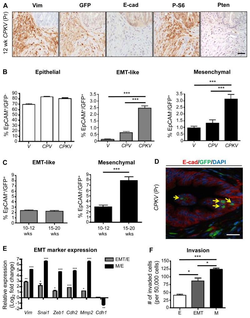
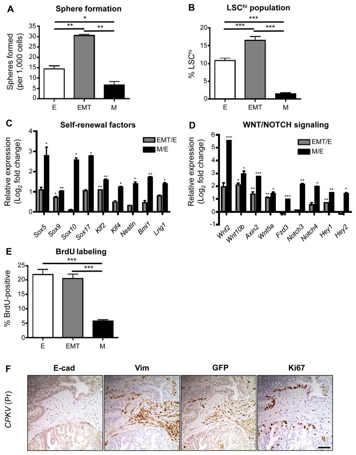
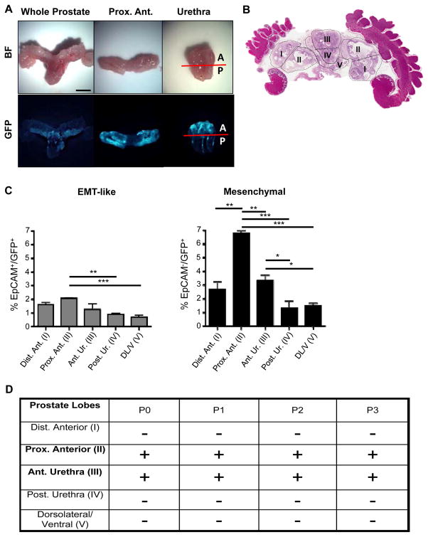
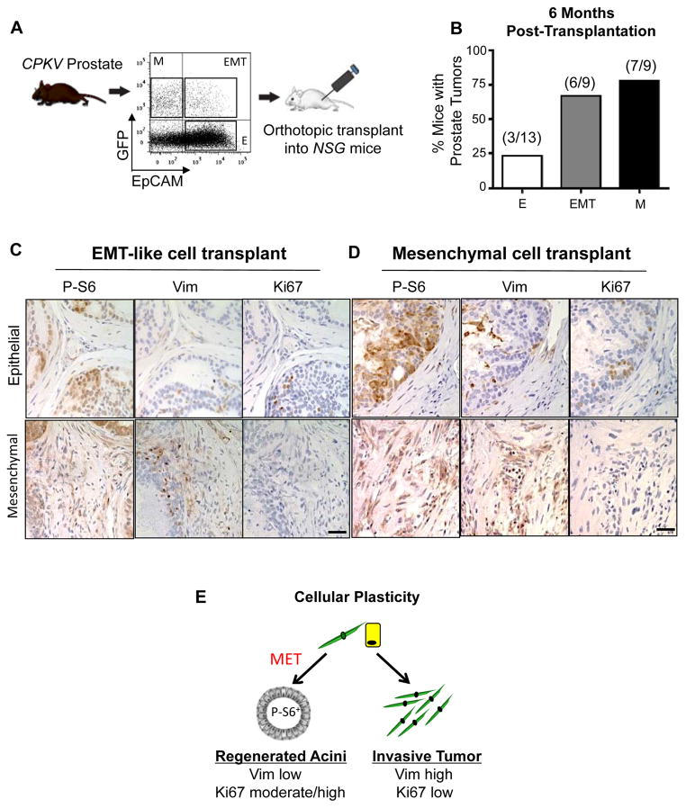
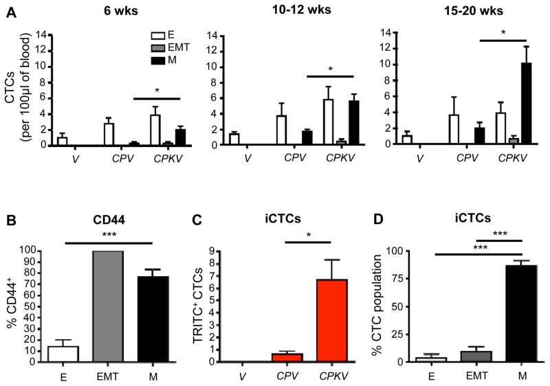
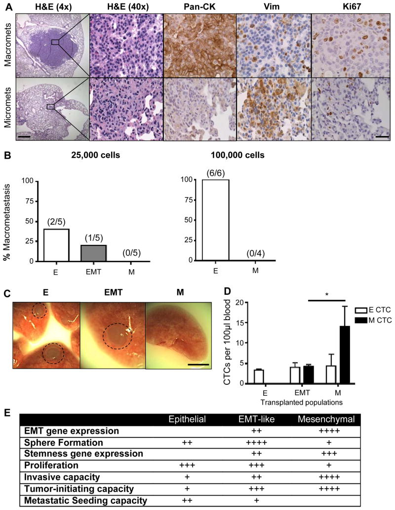
The epithelial-mesenchymal transition (EMT) has been postulated as a mechanism by which cancer cells acquire the invasive and stem-like traits necessary for distant metastasis. However, direct in vivo evidence for the role of EMT in the formation of cancer stem-like cells (CSC) and the metastatic cascade remains lacking. Here we report the first isolation and characterization of mesenchymal-like and EMT tumor cells, which harbor both epithelial and mesenchymal characteristics, in an autochthonous murine model of prostate cancer. By crossing the established Pb-Cre(+/-);Pten(L/L);Kras(G12D) (/+) prostate cancer model with a vimentin-GFP reporter strain, generating CPKV mice, we were able to isolate epithelial, EMT, and mesenchymal-like cancer cells based on expression of vimentin and EpCAM. CPKV mice (but not mice with Pten deletion alone) exhibited expansion of cells with EMT (EpCAM(+)/Vim-GFP(+)) and mesenchymal-like (EpCAM(-)/Vim-GFP(+)) characteristics at the primary tumor site and in circulation. These EMT and mesenchymal-like tumor cells displayed enhanced stemness and invasive character compared with epithelial tumor cells. Moreover, they displayed an enriched tumor-initiating capacity and could regenerate epithelial glandular structures in vivo, indicative of epithelia-mesenchyme plasticity. Interestingly, while mesenchymal-like tumor cells could persist in circulation and survive in the lung following intravenous injection, only epithelial and EMT tumor cells could form macrometastases. Our work extends the evidence that mesenchymal and epithelial states in cancer cells contribute differentially to their capacities for tumor initiation and metastatic seeding, respectively, and that EMT tumor cells exist with plasticity that can contribute to multiple stages of the metastatic cascade.


Conflict of interest statement

Disclosure of Potential Conflicts of Interest: The authors do not have potential conflicts of interest.

Figures

Figure 1
Figure 1
Tracking EMT and mesenchymal tumor cells in an endogenous prostate cancer model using a Vimentin-GFP reporter line. A, Prostates from CPKV mice (12 weeks) display EMT regions that are Vimentin (Vim)/GFP-positive surrounding E-cadherin (E-cad)-positive epithelial glandular structures. P-S6 marks cells that have undergone Cre recombination. B, Vimentin-GFP (GFP) and EpCAM were used to characterize epithelial (EpCAM+/GFP), EMT (EpCAM+/GFP+), and mesenchymal (EpCAM/GFP+) cell populations from the prostates of various Vim-GFP reporter mice (10–12 weeks) by FACS. CPKV mice have significant expansion of EMT and mesenchymal tumor cell populations. C, Expansion of mesenchymal tumor cell populations in CPKV prostates during late stage tumor progression (15–20 weeks). D, EMT cells (yellow arrow) that are GFP+ (green) and E-cadherin (E-cad)+ (red) are found within epithelial glandular structures in CPKV prostates (12 weeks). E, qPCR analysis confirms EMT signature gene expression in EMT and mesenchymal tumor cells isolated from CPKV prostates (10–12 weeks). F, Matrigel invasion assay reveals that EMT and mesenchymal tumor cells isolated from CPKV prostates (10–12 weeks) are significantly more invasive than epithelial tumor cells. Data in B, C, E, and F are represented as mean ± SEM. Bar, 50 μm. Pr, prostate. Lin,CD45/CD31/Ter119. *, P < 0.05, **, P < 0.01. ***, P < 0.001.
Figure 2
Figure 2
EMT and mesenchymal tumor cells have enhanced stemness properties. A, Matrigel sphere assay reveals that EMT tumor cells sorted from CPKV prostates (10–12 weeks) form more spheres than epithelial and mesenchymal tumor cells after 7 days in culture. B, EMT tumor cells have a higher LSChi content compared to epithelial and mesenchymal tumor cells from CPKV prostates (10–12 weeks), as quantified by FACS. C, EMT and mesenchymal tumor cells from CPKV prostates (10–12 weeks) have enhanced expression of self-renewal and stemness factors compared to epithelial cells. D, EMT and mesenchymal tumor cells from CPKV prostates (10–12 weeks) have enhanced expression of genes involved in WNT and NOTCH signaling compared to epithelial cells. E, Epithelial and EMT tumor cells from CPKV prostates (10–12 weeks) have a higher percentage of cells in S-phase compared to mesenchymal tumor cells, as measured by the percentage of BrdU+ cells. F, Ki67-positive cells are found preferentially in E-cadherin (E-cad)-positive glandular structures compared to Vimentin (Vim)/GFP-positive EMT regions in the stroma of CPKV prostates (12 weeks). Data in A through E are represented as mean ± SEM. Bar, 100 μm. Pr, prostate. *, P < 0.05, **, P < 0.01. ***, P < 0.001.
Figure 3
Figure 3
Prostate regions enriched in Vim-GFP+ cells are able to regenerate transplantable tumors in vivo. A, Bright-field (BF) and fluorescent images of a whole-mount CPKV prostate (10 weeks). GFP expression in CPKV prostates is most prominent in the proximal region of the anterior lobes and in the anterior portion of the urethra. B, Diagram depicting how prostate regions/lobes were separated. I, distal region of the anterior lobe. II, proximal region of the anterior lobe. III, anterior portion of the urethra. IV, posterior portion of the urethra. V, dorsolateral/ventral lobes. C, FACS analysis demonstrates that the anterior portion of the urethra and proximal region of the anterior lobes express the highest percentage of mesenchymal cells compared to other regions/lobes from the prostates of CPKV mice (10–12 weeks). The percentage of EMT tumor cells was also slightly higher in the proximal region of the anterior lobes compared to other lobes/regions. D, The only CPKV prostate regions/lobes that produced transplantable tumors in NSG mice were the proximal region of the anterior lobe and anterior portion of the urethra. Data in C are represented as mean ± SEM. Bar, 4mm. A, anterior. P, posterior. Dist. Ant., distal region of the anterior lobe. Prox. Ant., proximal region of the anterior lobe. Ant. Ur., anterior portion of the urethra. Post. Ur., posterior portion of the urethra. DL/V, dorsolateral/ventral lobes. P0, passage 0. P1, passage 1. P2, passage 2. P3, passage 3. +, tumor. −, no tumor. *, P < 0.05, **, P < 0.01. ***, P < 0.001.
Figure 4
Figure 4
EMT and mesenchymal tumor cells have enhanced tumor-initiating capacity and cellular plasticity in vivo. A, Schematic of experimental design for orthotopic transplantations. B, EMT and mesenchymal tumor cells form tumors more readily in vivo compared to epithelial tumor cells from CPKV prostates (10–12 weeks). C, IHC stains of anterior lobes from an NSG mouse transplanted with EMT tumor cells from CPKV prostates (10–12 weeks). P-S6 was used to trace transplanted cells, and Vim was used to mark mesenchymal cells. Top panel, transplanted EMT tumor cells form regenerated glandular structures. Bottom panel, transplanted EMT tumor cells form invasive mesenchymal tumor regions. D, Same as in C, except a prostate section from an NSG mouse transplanted with mesenchymal tumor cells from CPKV prostates (10–12 weeks). E, EMT (yellow) and mesenchymal (green) tumor cells from CPKV prostates have the plasticity to undergo an MET and regenerate epithelial glandular structures, or form invasive mesenchymal tumors in vivo. Bar, 50 μm.
Figure 5
Figure 5
Increase in CTCs with mesenchymal and invasive characteristics during disease progression in CPKV mice. A, CPKV mice have a significant increase in mesenchymal and EMT CTCs compared to CPV or V mice during disease progression. B, Mesenchymal and EMT CTCs from CPKV mice (15–20 weeks) have significantly higher CD44 expression compared to epithelial CTCs. C, CPKV mice have significantly more invasive CTCs (iCTCs) compared to CPV and V mice (15–20 weeks). D, The majority of iCTCs have a mesenchymal phenotype. Data in A through D are represented as mean ± SEM. *, P < 0.05, ***, P < 0.001.
Figure 6
Figure 6
Epithelial tumor cells have enhanced metastatic seeding potential. A, IHC analysis of epithelial (Pan-CK), mesenchymal (Vim), and proliferation (Ki67) markers in micrometastases (micromets) and macrometastases (macromets) in primary CPKV lungs (18 weeks). Low magnification bar, 500 μm; high magnification bar, 50 μm. B, Percent of macrometastatic lesions in the lungs of NSG mice 16 weeks after intravenous transplantation of either 25,000 or 100,000 epithelial, EMT, or mesenchymal tumor cells from CPKV prostates (10–12 weeks). C, Whole-mount images of lungs isolated from NSG mice transplanted with 25,000 epithelial, EMT, or mesenchymal tumor cells from CPKV prostates (10–12 weeks). Circle, macrometastases. Bar, 4 mm. D, NSG mice transplanted intravenously with mesenchymal tumor cells (25,000) from CPKV prostates (10–12 weeks) contained a significantly higher number of CTCs persisting in the bloodstream compared to mice transplanted with either epithelial or EMT tumor cells 16 weeks post-transplantation. E, Table summarizing the characteristics of epithelial, EMT, and mesenchymal tumor cells isolated from CPKV prostates. Data in D are represented as mean ± SEM. *, P < 0.05

References

    1. Siegel R, Ma J, Zou Z, Jemal A. Cancer statistics, 2014. CA: a cancer journal for clinicians. 2014;64:9–29. - PubMed
    1. Heidenreich A, Pfister D, Merseburger A, Bartsch G. Castration-resistant prostate cancer: where we stand in 2013 and what urologists should know. European urology. 2013;64:260–5. - PubMed
    1. Taylor BS, Schultz N, Hieronymus H, Gopalan A, Xiao Y, Carver BS, et al. Integrative genomic profiling of human prostate cancer. Cancer cell. 2010;18:11–22. - PMC - PubMed
    1. Mulholland DJ, Kobayashi N, Ruscetti M, Zhi A, Tran LM, Huang J, et al. Pten loss and RAS/MAPK activation cooperate to promote EMT and metastasis initiated from prostate cancer stem/progenitor cells. Cancer research. 2012;72:1878–89. - PMC - PubMed
    1. Aytes A, Mitrofanova A, Kinkade CW, Lefebvre C, Lei M, Phelan V, et al. ETV4 promotes metastasis in response to activation of PI3-kinase and Ras signaling in a mouse model of advanced prostate cancer. Proceedings of the National Academy of Sciences of the United States of America. 2013;110:E3506–15. - PMC - PubMed

Publication types