. 2015 Jun 15;194(12):6024-34.
doi: 10.4049/jimmunol.1500124. Epub 2015 May 6.

Regulation of neutrophilic inflammation by proteinase-activated receptor 1 during bacterial pulmonary infection

Affiliations

Regulation of neutrophilic inflammation by proteinase-activated receptor 1 during bacterial pulmonary infection

Ricardo J José et al. J Immunol. .

Abstract

Neutrophils are key effector cells of the innate immune response to pathogenic bacteria, but excessive neutrophilic inflammation can be associated with bystander tissue damage. The mechanisms responsible for neutrophil recruitment to the lungs during bacterial pneumonia are poorly defined. In this study, we focus on the potential role of the major high-affinity thrombin receptor, proteinase-activated receptor 1 (PAR-1), during the development of pneumonia to the common lung pathogen Streptococcus pneumoniae. Our studies demonstrate that neutrophils were indispensable for controlling S. pneumoniae outgrowth but contributed to alveolar barrier disruption. We further report that intra-alveolar coagulation (bronchoalveolar lavage fluid thrombin-antithrombin complex levels) and PAR-1 immunostaining were increased in this model of bacterial lung infection. Functional studies using the most clinically advanced PAR-1 antagonist, SCH530348, revealed a key contribution for PAR-1 signaling in influencing neutrophil recruitment to lung airspaces in response to both an invasive and noninvasive strain of S. pneumoniae (D39 and EF3030) but that PAR-1 antagonism did not impair the ability of the host to control bacterial outgrowth. PAR-1 antagonist treatment significantly decreased pulmonary levels of IL-1β, CXCL1, CCL2, and CCL7 and attenuated alveolar leak. Ab neutralization studies further demonstrated a nonredundant role for IL-1β, CXCL1, and CCL7 in mediating neutrophil recruitment in response to S. pneumoniae infection. Taken together, these data demonstrate a key role for PAR-1 during S. pneumoniae lung infection that is mediated, at least in part, by influencing multiple downstream inflammatory mediators.


Figures

FIGURE 1.
FIGURE 1.
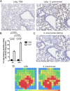
Characterization of neutrophil recruitment and S. pneumoniae infection in BALB/c mice. BALB/c mice (7–8 wk old) were challenged intranasally with S. pneumoniae D39 (5 × 10⁶ CFU) or PBS and culled at 4 h and 24 h for (A) immunohistochemistry of lung sections at 24 h stained with a specific Ly6G Ab and counterstained with hematoxylin. Original magnification ×10; inset, ×40. (B) Quantification of Ly6G staining at 4 and 24 h on lung sections from PBS-challenged and S. pneumoniae–infected mice. (C) Immunohistochemistry for S. pneumoniae serotype 2 on lung sections at 24 h from mice challenged with S. pneumoniae (original magnification ×10; inset, ×40, counterstained with hematoxylin) and (D) heat maps representative of Ly6G and S. pneumoniae staining intensity at 24 h. Data are expressed as bar graphs with means ± SEM (n = 6/group) and analyzed with the unpaired Student t test and two-way ANOVA with a Holm–Sidak post hoc test.
FIGURE 2.
FIGURE 2.
Neutrophil depletion protects mice from alveolar leak but compromises host defense. BALB/c mice (7–8 wk old) were treated with anti-Ly6G or anti-IgG (control Ab) 24 h prior to being challenged intranasally with S. pneumoniae D39 (5 × 10⁶ CFU). Mice were culled at 24 h postinfection. (A) Representative cytospins of cells recovered from BAL fluid (Romanowski stain; original magnification ×40) and (B) neutrophil counts. (C) Albumin levels recovered from BAL fluid 24 h following infection were measured as a marker of alveolar leak. Bacterial CFU recovered from (D) BAL fluid, (E) lung homogenates, and (F) blood were measured 24 h following infection.
FIGURE 3.
FIGURE 3.
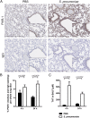
PAR-1 expression and coagulation activation is increased following S. pneumoniae infection. BALB/c mice (7–8 wk old) were challenged intranasally with S. pneumoniae D39 (5 × 10⁶ CFU) or PBS and culled at 4 h or 24 h. (A) Immunohistochemistry staining for PAR-1 on lung sections from mice challenged with PBS and S. pneumoniae 24 h postinfection (original magnification ×10; inset, ×40; IgG, negative control; counterstained with hematoxylin). (B) Quantification of PAR-1 staining intensity in lung sections at 4 and 24 h for mice challenged with PBS or infected with S. pneumoniae. (C) TAT complexes were measured by ELISA. Data are expressed as bar graphs with means ± SEM (n = 3–6/group) and analyzed with two-way ANOVA with a Holm–Sidak post hoc test.
FIGURE 4.
FIGURE 4.
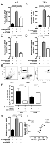
Neutrophil recruitment to the bronchoalveolar space and alveolar barrier disruption are attenuated by PAR-1 antagonism following S. pneumoniae infection. BALB/c mice (7–8 wk old) were challenged intranasally with PBS or S. pneumoniae D39 (5 × 10⁶ CFU) and treated with either vehicle or the PAR-1 antagonist. Total cells recovered from BAL fluid at (A) 4 and (B) 24 h following infection and the total number of neutrophils recovered from BAL fluid were calculated (C) 4 and (D) 24 h following infection. (E) Representative flow cytometry gating for the identification of neutrophils (Ly6GhiF4/80CD11bhi) and (F) proportion of neutrophils identified in BAL fluid and whole-lung homogenates at 4 h following infection in mice treated with the PAR-1 antagonist or vehicle. Furthermore, (G) BAL fluid albumin levels were measured by ELISA from recovered BAL fluid at 24 h postchallenge. Data are expressed as bar graphs with means ± SEM (n = 4–6/group) and were analyzed with one-way ANOVA with a Holm–Sidak post hoc test. Scatter plot is shown with linear regression line for neutrophil counts and albumin levels in (H) BAL fluid at 24 h for mice challenged with S. pneumoniae and treated with vehicle or the PAR-1 antagonist; data were analyzed with a Pearson correlation coefficient and linear regression.
FIGURE 5.
FIGURE 5.
PAR-1 antagonism reduces inflammation in S. pneumoniae infection. BALB/c mice (7–8 wk old) were challenged intranasally with PBS and or S. pneumoniae D39 (5 × 10⁶ CFU), treated with either vehicle or the PAR-1 antagonist, and culled at 4 h. (AF) Whole-lung homogenate levels of TNF, IL-6, IL-1β, IL-10, IFN-γ, and CXCL1 measured by multiplex ELISA (Meso Scale Discovery) and (G and H) whole-lung homogenate levels of CCL2 and CCL7 measured by ELISA. Data are expressed as means ± SEM (n = 4–6/group). Data were analyzed with one-way ANOVA with a Holm–Sidak post hoc test.
FIGURE 6.
FIGURE 6.
Neutralization of broncholaveolar CCL7 and IL-1β in the alveolar compartment and systemic neutralization of anti-CXCL1 reduce neutrophil recruitment to the bronchoalveolar space. BALB/c mice (7–8 wk old) were challenged intranasally with 10 μg neutralizing Ab (anti–IL-1β, anti-CCL7, anti-CCL2, and anti-CXCL1) and PBS or S. pneumoniae D39 (5 × 10⁶ CFU) and culled at 4 h. (AD) BAL fluid was recovered and the levels of IL-1β, CCL7, CCL2, and CXCL1 were measured. Neutrophil counts in BAL fluid were determined from cytospin preparations and total cells counts for mice treated intranasally with (E) anti–IL-1β, (F) anti-CCL7, (G) anti-CCL2, and (H) anti-CXCL1 and from mice treated with (I) anti-CXCL1 by i.p. injection. Data are expressed as bar graphs with means ± SEM (n = 5–6/group) and were analyzed with two-way ANOVA with a Holm–Sidak post hoc test.
FIGURE 7.
FIGURE 7.
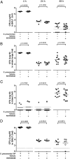
PAR-1 antagonism with SCH530348 does not compromise host defense. BALB/c mice (7–8 wk old) were challenged intranasally with PBS and treated with vehicle or challenged with S. pneumoniae D39 or S. pneumoniae EF3030 (5 × 10⁶ CFU) and treated with either vehicle or the PAR-1 antagonist. Mice were culled at 4, 24, or 48 h. Bacteria were cultured from recovered (A) BAL fluid, (B) whole-lung homogenates, and (C) blood at 4, 24, and 48 h, respectively, for mice challenged with D39 and (D) BAL fluid at 4, 24, and 48 h for mice challenged with EF3030. Data are expressed as dot plots with mean (n = 6–8/group) and analyzed with a Student t test.

Similar articles

Cited by

References

    1. Mizgerd J. P. 2006. Lung infection—a public health priority. PLoS Med. 3: e76 Available at: http://journals.plos.org/plosmedicine/article?id=10.1371/journal.pmed.00.... - PMC - PubMed
    1. Armstrong G. L., Conn L. A., Pinner R. W. 1999. Trends in infectious disease mortality in the United States during the 20th century. JAMA 281: 61–66. - PubMed
    1. World Health Organisation 2007. Pneumococcal conjugate vaccine for childhood immunization—WHO position paper. Wkly. Epidemiol. Rec. 82: 93–104. - PubMed
    1. Calbo E., Alsina M., Rodríguez-Carballeira M., Lite J., Garau J. 2010. The impact of time on the systemic inflammatory response in pneumococcal pneumonia. Eur. Respir. J. 35: 614–618. - PubMed
    1. Choi G., Hofstra J.-J. H., Roelofs J. J. T. H., Rijneveld A. W., Bresser P., van der Zee J. S., Florquin S., van der Poll T., Levi M., Schultz M. J. 2008. Antithrombin inhibits bronchoalveolar activation of coagulation and limits lung injury during Streptococcus pneumoniae pneumonia in rats. Crit. Care Med. 36: 204–210. - PubMed

Publication types

MeSH terms

LinkOut - more resources