

Solitary bone cysts-A rare occurrence with bilaterally symmetrical presentation
- PMID: 25949013
- PMCID: PMC4409203
- DOI: 10.4103/0973-029X.151366
Solitary bone cysts-A rare occurrence with bilaterally symmetrical presentation
Abstract
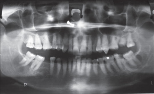
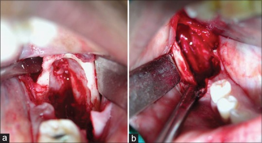
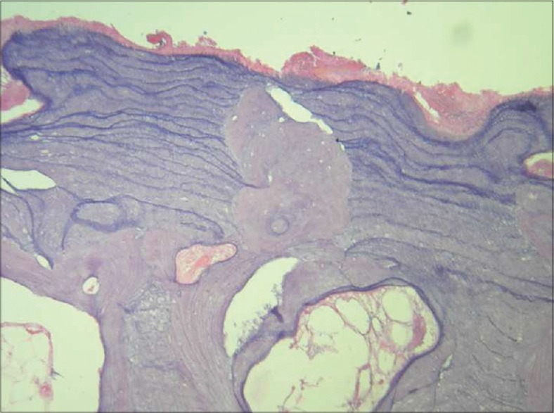
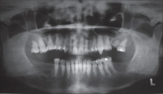
Solitary bone cysts (SBCs) are bone cavities that lack a true epithelial lining. They are more commonly seen during the first 2decades of age.Very few cases have been reported over 40 years of age.SBCs are usually discovered as an accidental coexisting finding during a routine radiologic examination or during another unrelated dental complaint. They present as a unilocular or multilocular radiolucent lesion associated with vital teeth with mild or no cortical expansion. Bilateral presentation is however very rare. We present a case of 52-year-old female patient with bilateral presentation of SBCs.
Keywords: Solitary bone cyst; simple bone cyst; traumatic bone cyst.
Conflict of interest statement
Figures
References
-
- Regezi JA, Scuibba JJ, Jordan RC. 4th ed. St Louis: Saunders; 2003. Oral Pathology-Clinical pathologic correlations; p. 259.
-
- Harnet JC, Lombardi T, Kiewansky P, Rieger J, Tempe MH, Clavert JM. Solitary bone cyst of the jaws: A review of etiopathogenic hypotheses. J Oral MaxillofacSurg. 2008;66:2345–8. - PubMed
-
- Suie Y, Taguchi A, Tanimato K. A comparative study of the simple bone cysts of the jaw and the extracranialbones. DentomaxillofacRadiol. 2007;36:125–9. - PubMed
-
- Sandev S, Sokler K, Grgurevic J. Traumatic bone cysts. ActaStomatol Croat. 2001;35:417–20.
Publication types
LinkOut - more resources
Full Text Sources
Other Literature Sources
