

Elliptical Morphology of the Carpal Tunnel Cross Section
- PMID: 25949095
- PMCID: PMC4418467
Elliptical Morphology of the Carpal Tunnel Cross Section
Abstract
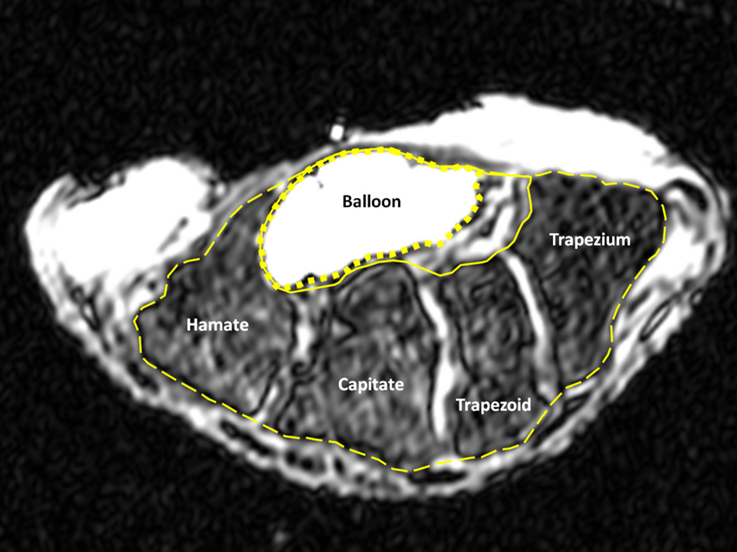
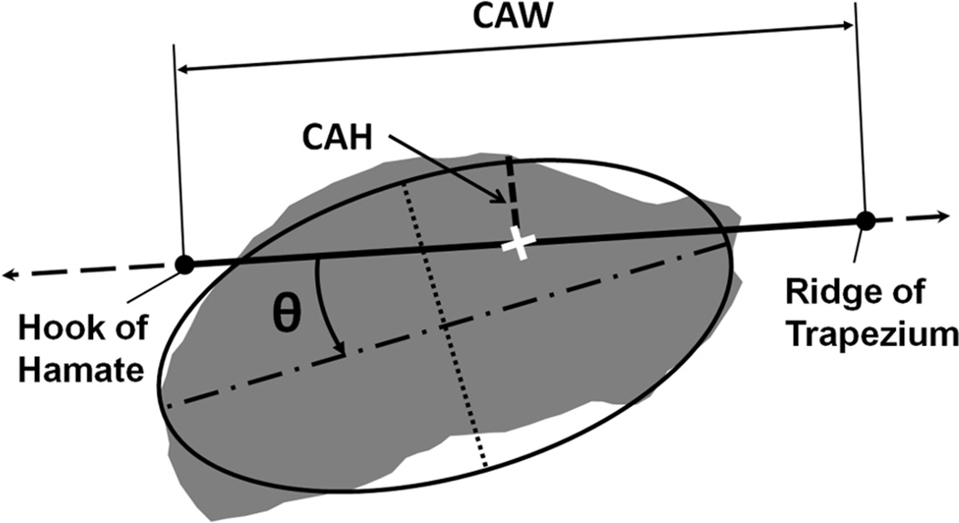
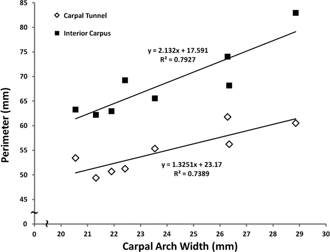
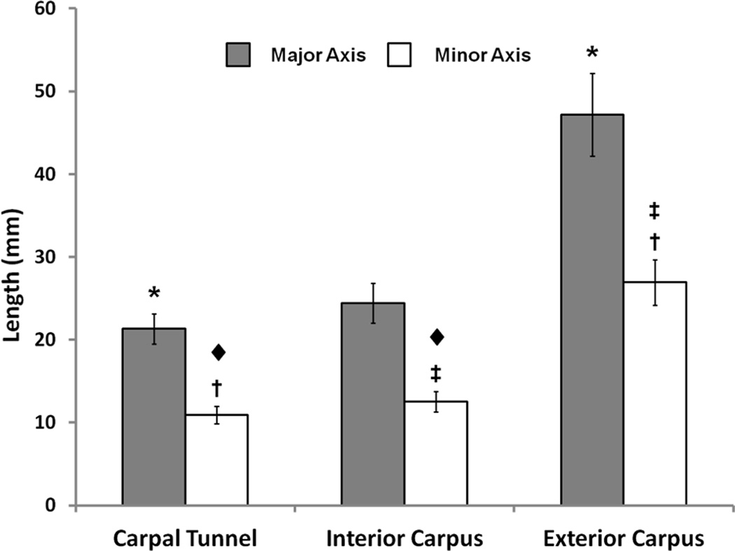
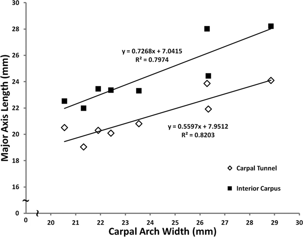
Although the carpal tunnel is known for its anatomical constituents, its morphology is not well recognized. The aim of this study was to investigate the morphometric properties of the carpal tunnel and its surrounding structures. Magnetic resonance, cross-sectional images of the distal carpal tunnel were collected from eight cadaveric hands. Morphological analyses were performed for the cross sections of the carpal tunnel, interior carpus boundary, and exterior carpus boundary. The specimens had a carpal arch width and height of 23.9 ± 2.9 mm and 2.2 ± 0.9 mm, respectively. The carpal tunnel, interior carpus boundary, and exterior carpus boundary had perimeters of 54.8 ± 4.5 mm, 68.5 ± 7.0 mm, and 130.6 ± 11.8 mm, respectively, and areas of 183.5 ± 30.1 mm2, 240.7 ± 40.2 mm2, and 1002.3 ± 183.7 mm2, respectively. The cross sections were characterized by elliptical fitting with aspect ratios of 1.96 ± 0.15, 1.96 ± 0.19, and 1.76 ± 0.19 for the carpal tunnel, interior carpus boundary, and exterior carpus boundary, respectively. The major axis of the boundaries increased in pronation angle, relative to the hamate-trapezium axis, for the exterior carpus (6.0 ± 3.0°), interior carpus (8.2 ± 3.2°), and carpal tunnel (15.9 ± 2.2°). This study advances our understanding of the structural anatomy of the carpal tunnel, and the morphological information is valuable in the identification of structural abnormality, assistance of surgical planning, and evaluation of treatment of effects.
Keywords: carpal tunnel; carpus; morphology.
Figures
Similar articles
-
Carpal Tunnel Cross-Sectional Area Affected by Soft Tissues Abutting the Carpal Bones.J Wrist Surg. 2013 Feb;2(102):73-78. doi: 10.1055/s-0032-1329593. J Wrist Surg. 2013. PMID: 23607081 Free PMC article.
-
Three-Dimensional Carpal Kinematics after Carpal Tunnel Release.J Wrist Surg. 2016 Aug;5(3):222-6. doi: 10.1055/s-0036-1578812. Epub 2016 Feb 19. J Wrist Surg. 2016. PMID: 27468373 Free PMC article.
-
Morphological analysis of the carpal tunnel.Hand (N Y). 2010 Mar;5(1):77-81. doi: 10.1007/s11552-009-9220-9. Epub 2009 Sep 4. Hand (N Y). 2010. PMID: 19760464 Free PMC article.
-
Practical anatomy of the carpal tunnel.Hand Clin. 2002 May;18(2):219-30. doi: 10.1016/s0749-0712(01)00003-8. Hand Clin. 2002. PMID: 12371025 Review.
-
Biomechanical and anatomical consequences of carpal tunnel release.Clin Biomech (Bristol). 2003 Oct;18(8):685-93. doi: 10.1016/s0268-0033(03)00052-4. Clin Biomech (Bristol). 2003. PMID: 12957554 Review.
Cited by
-
Mandibular Retromolar Foramen and Canal - A Systematic Review and Meta-Analysis.Ann Maxillofac Surg. 2020 Jul-Dec;10(2):444-449. doi: 10.4103/ams.ams_19_20. Epub 2020 Jun 18. Ann Maxillofac Surg. 2020. PMID: 33708593 Free PMC article. Review.
-
Comparing the Carpal Tunnel Area and Carpal Boundaries in Patients with Carpal Tunnel Syndrome and Healthy Volunteers: A Magnetic Resonance Imaging Study.Diagnostics (Basel). 2025 May 9;15(10):1205. doi: 10.3390/diagnostics15101205. Diagnostics (Basel). 2025. PMID: 40428198 Free PMC article.
-
Three-dimensional stiffness of the carpal arch.J Biomech. 2016 Jan 4;49(1):53-59. doi: 10.1016/j.jbiomech.2015.11.005. Epub 2015 Nov 18. J Biomech. 2016. PMID: 26617368 Free PMC article.
-
External wrist ratio is not a proxy for internal carpal tunnel shape: Implications for evaluating carpal tunnel syndrome risk.Clin Anat. 2024 Nov;37(8):869-877. doi: 10.1002/ca.24132. Epub 2024 Jan 3. Clin Anat. 2024. PMID: 38173294
-
Tendon of Flexor Carpi Radialis in carpal tunnel: a radiologic and cadaveric study.Turk J Med Sci. 2021 Aug 30;51(4):1912-1916. doi: 10.3906/sag-2012-31. Turk J Med Sci. 2021. PMID: 33705637 Free PMC article.
References
-
- Ablove RH, Peimer CA, Diao E, Oliverio R, Kuhn JP. Morphologic changes following endoscopic and two-portal subcutaneous carpal tunnel release. J Hand Surg Am. 1994;19:821–826. - PubMed
-
- Allmann KH, Horch R, Uhl M, Gufler H, Altehoefer C, Stark GB, Langer M. MR imaging of the carpal tunnel. Eur J Radiol. 1997;25:141–145. - PubMed
-
- Bleecker ML, Bohlman M, Moreland R, Tipton A. Carpal tunnel syndrome: role of carpal canal size. Neurology. 1985;35:1599–1604. - PubMed
-
- Brahme SK, Hodler J, Braun RM, Sebrechts C, Jackson W, Resnick D. Dynamic MR imaging of carpal tunnel syndrome. Skeletal Radiol. 1997;26:482–487. - PubMed
-
- Buchberger W, Schon G, Strasser K, Jungwirth W. High-resolution ultrasonography of the carpal tunnel. J Ultrasound Med. 1991;10:531–537. - PubMed
Grants and funding
LinkOut - more resources
Full Text Sources