. 2012 Dec;16(4):253-64.
doi: 10.12717/DR.2012.16.4.253.

Nesfatin-1 as a new potent regulator in reproductive system

Affiliations

Nesfatin-1 as a new potent regulator in reproductive system

Jinhee Kim et al. Dev Reprod. 2012 Dec.

Abstract

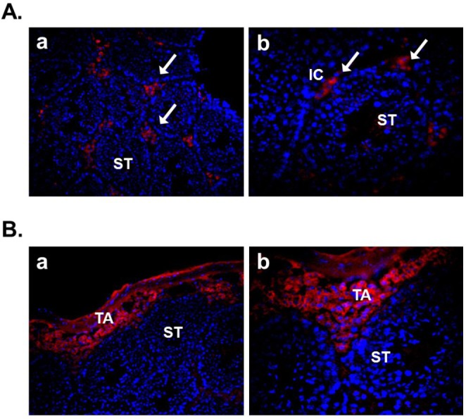
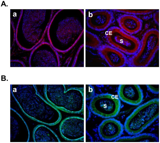
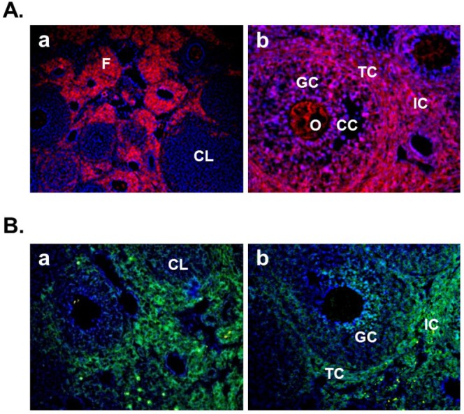
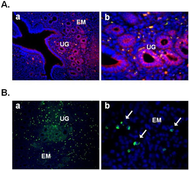
Nesfatin-1 is a recently discovered anorexigenic peptide which is distributed in several brain areas implicated in the feeding and metabolic regulation. Recently, it has been reported that nesfatin-1 is expressed not only in brain, but also in peripheral organs such as digestive organs, adipose tissues, heart, and reproductive organs. Nesfatin-1 is markedly expressed in the pancreas, stomach and duodenum. Eventually, the nesfatin-1 expression in the digestive organs may be regulated by nutritional status, which suggests a regulatory role of peripheral nesfatin-1 in energy homeostasis. Nesfatin-1 is also detected in the adipose tissues of humans and rodents, indicating that nesfatin-1 expression in the fat may regulate food intake independently, rather than relying on leptin. In addition, nesfatin-1 is expressed in the heart as a cardiac peptide. It suggests that nesfatin-1 may regulate cardiac function and encourage clinical potential in the presence of nutrition-dependent physio-pathologic cardiovascular diseases. Currently, only a few studies demonstrate that nesfatin-1 is expressed in the reproductive system. However, it is not clear yet what function of nesfatin-1 is in the reproductive organs. Here, we summarize the expression of nesfatin-1 and its roles in brain and peripheral organs and discuss the possible roles of nesfatin-1 expressed in reproductive organs, including testis, epididymis, ovary, and uterus. We come to the conclusion that nesfatin-1 as a local regulator in male and female reproductive organs may regulate the steroidogenesis in the testis and ovary and the physiological activity in epididymis and uterus.

Keywords: Epididymis; Nesfatin-1; Ovary; Testis; Uterus.


Figures

Fig. 1.
Fig. 1.
The localization of nesfatin-1 protein and its binding sites in mouse testis. (A) Testis section was stained with anti-nesfatin-1 antibody to localize nesfatin-1. Nesfatin-1 was expressed within the interstitial cells, including the Leydig cells between the seminiferous tubules. a: 200×; b: 400×. (B) Testis section was stained with biotin-conjugated nesfatin-1 and Alexa flour 594-conjugated streptavidin to detect the nesfatin-1 binding sites. Nesfatin-1 binding sites were localized at boundary cells in tunica albuginea. a: 200×; b: 400×. (ST, seminiferous tubule; IC, interstitial cell; TA, tunica albuginea)
Fig. 2.
Fig. 2.
The localization of nesfatin-1 protein and its binding sites in mouse epididymis. (A) Epididymis section was stained with anti-nesfatin-1 antibody to localize nesfatin-1. Nesfatin-1 was expressed at the columnar epithelium and some of the spermatozoa in the lumen. a: 200×; b: 400×. (B) Epididymis section was stained with FITC-conjugated nesfatin-1 to detect the nesfatin-1 binding sites. Nesfatin-1 binding sites were localized at the columnar epithelium and some of the spermatozoa in the lumen. Interestingly, nesfatin-1 binding sites were detected at same location as nesfatin-1 expression sites. a: 200×; b: 400×. (S, spermatozoa; CE, columnar epithelium)
Fig. 3.
Fig. 3.
The localization of nesfatin-1 protein and its binding sites in mouse ovary. (A) Ovarian section was stained with anti-nesfatin-1 antibody to localize nesfatin-1 protein. Nesfatin-1 was expressed in the theca cells around the follicles, the interstitial cells, and the oocytes in some of the follicles. Interestingly, the granulosa cells in the some of the follicles and the cumulus cells around the oocytes were stained with nesfatin-1, but not the corpus luteum. a: 200×; b: 400×. (B) Ovarian section was stained with FITC-conjugated nesfatin-1 to detect the nesfatin-1 binding sites. Nesfatin-1 binding sites were localized in the theca cells and interstitial cells around follicles. Interestingly, some granulosa cells in the follicles were stained with FITC-conjugated nesfatin-1. a, b: 200×. (F, follicle; CL, corpus luteum; O, oocyte; GC, granulosa cells; CC, cumulus cells; IC, interstitial cells; TC, theca cells)
Fig. 4.
Fig. 4.
The localization of nesfatin-1 protein and its binding sites in mouse uterus. (A) Uterine section was stained with antinesfatin-1 antibody to localize nesfatin-1. Nesfatin-1 was expressed on the epithelial cells of endometrium and uterine glands. a: 100×; b: 400×. (B) Uterine section was stained with FITC-conjugated nesfatin-1 to detect the nesfatin-1 binding sites. Nesfatin-1 binding sites were localized in the epithelial cells of uterine glands. Interestingly, neutrophils in the endometrium were stained with FITC-conjugated nesfatin-1 (arrows in b). a: 100×; b: 1,000×. (EM, endometrium; UG, uterine gland)

Similar articles

Cited by

References

    1. Akingbemi BT, Sottas CM, Koulova AI, Klinefelter GR, Hardy MP. Inhibition of testicular steroidogenesis by the xenoestrogen bisphenol A is associated with reduced pituitary luteinizing hormone secretion and decreased steroidogenic enzyme gene expression in rat Leydig cells. Endocrinology. 2004;145:592–603. - PubMed
    1. Angelone T, Filice E, Pasqua T, Amodio N, Galluccio M, Montesanti G, Quintieri AM, Cerra MC. Nesfatin-1 as a novel cardiac peptide identification, functional characterization, and protection against ischemia/reperfusion injury. Cell Mol Life Sci. 2012. Epub ahead of print. - PMC - PubMed
    1. Aydin S, Dag E, Ozkan Y, Erman F, Dagli AF, Kilic N, Sahin I, Karatas F, Yoldas T, Barim AO, Kendir Y. Nesfatin-1 and ghrelin levels in serum and saliva of epileptic patients: hormonal changes can have a major effect on seizure disorders. Mol Cell Biochem. 2009;328:49–56. - PubMed
    1. Barnikol-Watanabe S, Gross NA, Gotz H, Henkel T, Karabinos A, Kratzin H, Barnikol HU, Hilschmann N. Human protein NEFA, a novel DNA binding/EF-hand/leucine zipper protein. Molecular cloning and sequence analysis of the cDNA, isolation and characterization of the protein. Biol Chem Hoppe Seyler. 1994;375:497–512. - PubMed
    1. Bonnet MS, Pecchi E, Trouslard J, Jean A, Dallaporta M, Troadec JD. Central nesfatin-1 expressing neurons are sensitive to peripheral inflammatory stimulus. J Neuroinflammation. 2009;6:27. - PMC - PubMed