The Expression Pattern of Melatonin Receptor 1a Gene during Early Life Stages in the Nile tilapia (Oreochromis niloticus)
- PMID: 25949120
- PMCID: PMC4282221
- DOI: 10.12717/DR.2013.17.1.045
The Expression Pattern of Melatonin Receptor 1a Gene during Early Life Stages in the Nile tilapia (Oreochromis niloticus)
Abstract
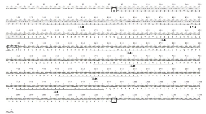
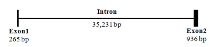
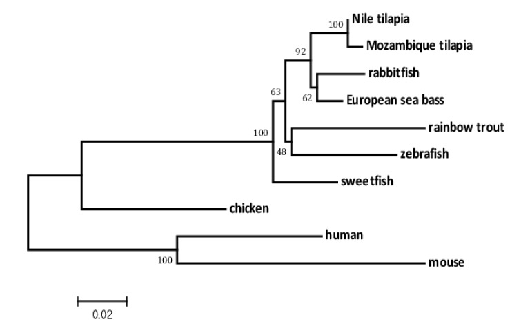
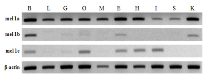
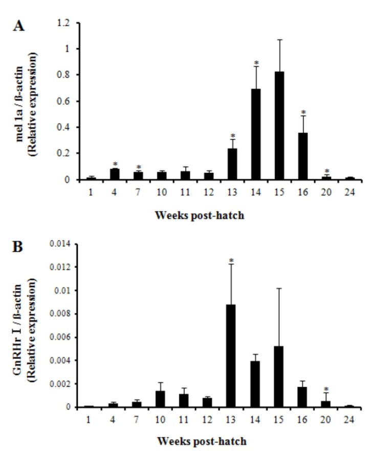
The action of melatonin within the body of animals is known to be mediated by melatonin receptors. Three different types of melatonin receptors have been identified so far in fish. However, which of these are specifically involved in puberty onset is not known in fish. We cloned and analyzed the sequence of melatonin receptor 1a (mel 1a) gene in Nile tilapia Oreochromis niloticus. In addition, we examined the tissue distribution of gene expressions for three types of receptors, mel 1a, 1b and lc and investigated which of them is involved in the onset of puberty by comparing their expression with that of gonadotropin-releasing hormone receptor I (GnRHr I) gene using quantitative real-time PCR from 1 week post hatch (wph) to 24 wph. The mel 1a gene of Nile tilapia consisted of two exons and one bulky intron between them. Mel 1a gene was found to be highly conserved gene showing high homology with the corresponding genes from different teleost. All three types of melatonin receptor genes were expressed in the brain, eyes and ovary in common. Expression of mel 1a gene was the most abundant and ubiquitous among 3 receptors in the brain, liver, gill, ovary, muscle, eye, heart, intestine, spleen and kidney. Mel 1b and mel 1c genes were, however, expressed in fewer tissues at low level. During the development post hatch, expressions of both mel 1a and GnRHr I genes significantly increased at 13 wph which was close to the putative timing of puberty onset in this species. These results suggest that among three types of receptors mel 1a is most likely associated with the action of melatonin in the onset of puberty in Nile tilapia.
Keywords: Mel 1a; Melatonin receptors; Nile tilapia; Oreochromis niloticus; Puberty.
Figures
Similar articles
-
Diurnal expressions of four subtypes of melatonin receptor genes in the optic tectum and retina of goldfish.Comp Biochem Physiol A Mol Integr Physiol. 2009 Feb;152(2):219-24. doi: 10.1016/j.cbpa.2008.09.030. Epub 2008 Oct 5. Comp Biochem Physiol A Mol Integr Physiol. 2009. PMID: 18930834
-
Expression analysis of melatonin receptor subtypes in the ovary of domestic chicken.Vet Res Commun. 2009 Jan;33(1):49-56. doi: 10.1007/s11259-008-9071-9. Epub 2008 Jul 5. Vet Res Commun. 2009. PMID: 18604592
-
Melatonin receptor genes (mel-1a, mel-1b, mel-1c) are differentially expressed in the avian germ line.Mol Reprod Dev. 2008 Sep;75(9):1408-17. doi: 10.1002/mrd.20885. Mol Reprod Dev. 2008. PMID: 18288645
-
Kisspeptin2 stimulates the HPG axis in immature Nile tilapia (Oreochromis niloticus).Comp Biochem Physiol B Biochem Mol Biol. 2016 Dec;202:31-38. doi: 10.1016/j.cbpb.2016.07.009. Epub 2016 Aug 3. Comp Biochem Physiol B Biochem Mol Biol. 2016. PMID: 27497664
-
Molecular characterization of the prolactin receptor in two fish species, tilapia Oreochromis niloticus and rainbow trout, Oncorhynchus mykiss: a comparative approach.Can J Physiol Pharmacol. 2000 Dec;78(12):1086-96. Can J Physiol Pharmacol. 2000. PMID: 11149385 Review.
Cited by
-
Contribution of opsins and chromophores to cone pigment variation across populations of Lake Victoria cichlids.J Fish Biol. 2022 Aug;101(2):365-377. doi: 10.1111/jfb.14969. Epub 2021 Dec 29. J Fish Biol. 2022. PMID: 34860424 Free PMC article.
-
Phylogenetic Reclassification of Vertebrate Melatonin Receptors To Include Mel1d.G3 (Bethesda). 2019 Oct 7;9(10):3225-3238. doi: 10.1534/g3.119.400170. G3 (Bethesda). 2019. PMID: 31416806 Free PMC article.
-
Seasonal expression of reproductive axis-related neuroendocrine genes and their relation with ovarian maturation in captive yellowtail kingfish (Seriola lalandi).Biol Res. 2025 Aug 8;58(1):55. doi: 10.1186/s40659-025-00622-5. Biol Res. 2025. PMID: 40775662 Free PMC article.
References
-
- Amano M, Iigo M, Ikuta K, Kitamura S, Okuzawa K, Yamada H, Yamamori K. Disturbance of plasma melatonin profile by high dose melatonin administration inhibits testicular maturation of precocious male masu salmon. Zool Sci. 2004;21:79–85. - PubMed
-
- Ballesteros JA, Jensen AD, Liapakis G, Rasmussen SG, Shi L, Gether U, Javitch JA. Activation of the beta 2-adrenergic receptor involves disruption of an ionic lock between the cytoplasmic ends of transmembrane segments 3 and 6. J Biol Chem. 2001;276:29171–29177. - PubMed
-
- Bayarri MJ, Falcón J, Zanuy S, Carrillo M. Continuous light and melatonin daily and seasonal variations of brain binding sites and plasma concentration during the first reproductive cycle of sea bass. Gen Comp Endocrinol. 2010;169:58–64. - PubMed
-
- Bayarri MJ, Rodriguez L, Zanuy S, Madrid JA, Sanchez- Vazquez FJ, Kagawa H, Okuzawa K, Carrillo M. Effect of photoperiod manipulation on the daily rhythms of melatonin and reproductive hormones in caged European sea bass (Dicentrarchus labrax) Gen Comp Endocrinol. 2004;136:72–81. - PubMed
-
- Bhattacharya S, Chattoraj A, Maitra SK. Melatonin in the regulation of annual testicular events in carp Catla catla evidence from the studies on the effects of exogenous melatonin, continuous light, and continuous darkness. Chronobiol Int. 2007;24:629–650. - PubMed
LinkOut - more resources
Full Text Sources
Other Literature Sources