PKCη Regulates the TGFβ3-induced Chondevrepogenic Differentiation of Human Mesenchymal Stem Cell
- PMID: 25949145
- PMCID: PMC4382954
- DOI: 10.12717/DR.2013.17.4.299
PKCη Regulates the TGFβ3-induced Chondevrepogenic Differentiation of Human Mesenchymal Stem Cell
Abstract
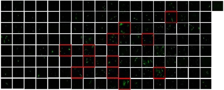
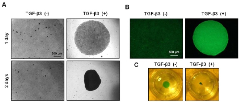
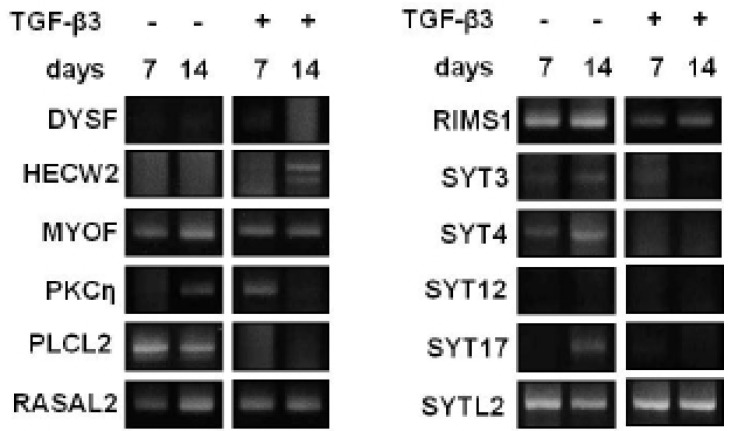
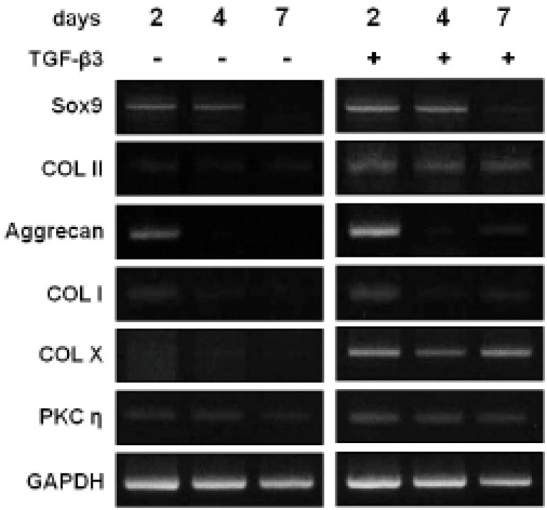
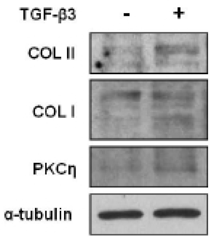
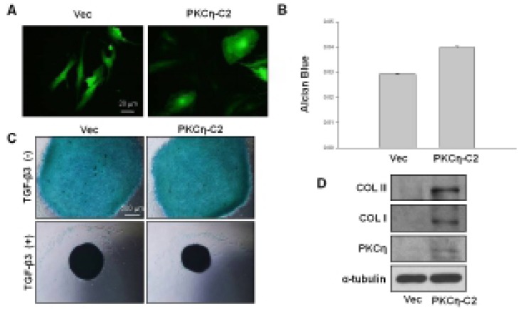
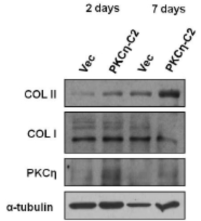
Transforming growth factor (TGF) family is well known to induce the chondevrepogenic differentiation of mesenchymal stem cells (MSC). However, the precise signal transduction pathways and underlying factors are not well known. Thus the present study aims to evaluate the possible role of C2 domain in the chondevrepogenic differentiation of human mesenchymal stem cells. To this end, 145 C2 domains in the adenovirus were individually transfected to hMSC, and morphological changes were examined. Among 145 C2 domains, C2 domain of protein kinase C eta (PKCη) was selected as a possible chondevrepogenic differentiation factor for hMSC. To confirm this possibility, we treated TGFβ3, a well known chondevrepogenic differentiation factor of hMSC, and examined the increased-expression of glycosaminoglycan (GAG), collagen type II (COL II) as well as PKCη using PT-PCR, immunocytochemistry and Western blot analysis. To further evaluation of C2 domain of PKCη, we examined morphological changes, expressions of GAG and COL II after transfection of PKCη -C2 domain in hMSC. Overexpression of PKCη-C2 domain induced morphological change and increased GAG and COL II expressions. The present results demonstrate that PKCη involves in the TGF-β3-induced chondevrepogenic differentiation of hMSC, and C2 domain of PKCη has important role in this process.
Keywords: C2-domain.; Chondevrepogenesis; Human mesenchymal stem cell; PKCη; TGF-β3.
Figures
Similar articles
-
The Enigmatic Protein Kinase C-eta.Cancers (Basel). 2019 Feb 13;11(2):214. doi: 10.3390/cancers11020214. Cancers (Basel). 2019. PMID: 30781807 Free PMC article. Review.
-
[CONSTRUCTION AND IDENTIFICATION OF ADENOVIRUS VECTOR EXPRESSING BONE MORPHOGENETIC PROTEIN 2 AND TRANSFORMING GROWTH FACTOR β3 GENES AND THEIR EXPRESSION IN BONE MARROW MESENCHYMAL STEM CELLS OF DIANNAN SMALL-EAR PIGS].Zhongguo Xiu Fu Chong Jian Wai Ke Za Zhi. 2014 Jul;28(7):896-902. Zhongguo Xiu Fu Chong Jian Wai Ke Za Zhi. 2014. PMID: 26462358 Chinese.
-
[Chondrogenesis of bone marrow mesenchymal stem cells induced by transforming growth factor beta3 gene in Diannan small-ear pigs].Zhongguo Xiu Fu Chong Jian Wai Ke Za Zhi. 2014 Feb;28(2):149-54. Zhongguo Xiu Fu Chong Jian Wai Ke Za Zhi. 2014. PMID: 24796184 Chinese.
-
Interplay between local versus soluble transforming growth factor-beta and fibrin scaffolds: role of cells and impact on human mesenchymal stem cell chondrogenesis.Tissue Eng Part A. 2012 Jun;18(11-12):1140-50. doi: 10.1089/ten.TEA.2011.0426. Epub 2012 May 14. Tissue Eng Part A. 2012. PMID: 22480213
-
Differentiation of MSC and annulus fibrosus cells on genetically engineered silk fleece-membrane-composites enriched for GDF-6 or TGF-β3.J Orthop Res. 2018 May;36(5):1324-1333. doi: 10.1002/jor.23778. Epub 2017 Nov 22. J Orthop Res. 2018. PMID: 29058815
Cited by
-
The HRAS-binding C2 domain of PLCη2 suppresses tumor-like synoviocytes and experimental arthritis in rheumatoid arthritis.Exp Mol Med. 2025 Feb;57(2):335-348. doi: 10.1038/s12276-025-01393-5. Epub 2025 Feb 3. Exp Mol Med. 2025. PMID: 39894825 Free PMC article.
-
The Enigmatic Protein Kinase C-eta.Cancers (Basel). 2019 Feb 13;11(2):214. doi: 10.3390/cancers11020214. Cancers (Basel). 2019. PMID: 30781807 Free PMC article. Review.
References
-
- Arita NA, Pelaez D, Cheung HS. Activation of the extracellular signal-regulated kinases 1 and 2 (ERK1/2) is needed for the TGFβ-induced chondevrepogenic and osteogenic differentiation of mesenchymal stem cells. Biochem Biophys Res Commun. 2011;405:564–569. - PubMed
-
- Augello A, De Bari C. The regulation of differentiation in mesenchymal stem cells. Hum Gnen Ther. 2010;21:1226–1238. - PubMed
-
- Bobick BE, Chen FH, Le AM, Tuan RS. Regulation of the chondevrepogenic phenotype in culture. Brith Defects Res CEmbryo Today. 2009;87:351–371. - PubMed
LinkOut - more resources
Full Text Sources
Other Literature Sources
Miscellaneous