Enhancement of Transgene Expression by HDAC Inhibitors in Mouse Embryonic Stem Cells
- PMID: 25949154
- PMCID: PMC4382945
- DOI: 10.12717/DR.2013.17.4.379
Enhancement of Transgene Expression by HDAC Inhibitors in Mouse Embryonic Stem Cells
Abstract
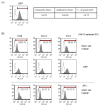
Embryonic stem (ES) cells can self-renew and differentiate to various cells depending on the culture condition. Although ES cells are a good model for cell type specification and can be useful for application in clinics in the future, studies on ES cells have many experimental restraints including low transfection efficiency and transgene expression. Here, we observed that transgene expression after transfection was enhanced by treatment with histone deacetylse (HDAC) inhibitors such as trichostatin A, sodium butyrate, and valproic acid. Transfection was performed using conventional transfection reagents with a retroviral vector encoding GFP under the control of CMV promoter as a reporter. Treatment of ES cells with HDAC inhibitors after transfection increased population of GFP positive cells up to 180% compared with untreated control. ES cells showed normal expression of stem cell markers after treatment with HDAC inhibitors. Transgene expression was further enhanced by modifying transfection procedure. GFP positive cells selected after transfection were proved to have the stem cell properties. Our improved protocol for enhanced gene delivery and expression in mouse ES cells without hampering ES cell properties will be useful for study and application of ES cells.
Keywords: Enhancement; HDAC inhibitor; Mouse embryonic stem cell; Optimization.; Transfection.
Figures


Similar articles
-
Histone deacetylase inhibitors preferentially augment transient transgene expression in human dermal fibroblasts.Br J Dermatol. 2007 Oct;157(4):662-9. doi: 10.1111/j.1365-2133.2007.08122.x. Epub 2007 Aug 17. Br J Dermatol. 2007. PMID: 17711521
-
Differentiation and upregulation of heat shock protein 70 induced by a subset of histone deacetylase inhibitors in mouse and human embryonic stem cells.BMB Rep. 2011 Mar;44(3):176-81. doi: 10.5483/BMBRep.2011.44.3.176. BMB Rep. 2011. PMID: 21429295
-
Efficiency of the elongation factor-1alpha promoter in mammalian embryonic stem cells using lentiviral gene delivery systems.Stem Cells Dev. 2007 Aug;16(4):537-45. doi: 10.1089/scd.2006.0088. Stem Cells Dev. 2007. PMID: 17784828
-
Histone deacetylase inhibitor promotes differentiation of embryonic stem cells into neural cells in adherent monoculture.Chin Med J (Engl). 2010 Mar 20;123(6):734-8. Chin Med J (Engl). 2010. PMID: 20368096
-
An Efficient Transfection Method for Differentiation and Cell Proliferation of Mouse Embryonic Stem Cells.Methods Mol Biol. 2017;1622:139-147. doi: 10.1007/978-1-4939-7108-4_11. Methods Mol Biol. 2017. PMID: 28674807
Cited by
-
Entinostat, a histone deacetylase inhibitor, enhances CAR-NK cell anti-tumor activity by sustaining CAR expression.Front Immunol. 2025 Mar 7;16:1533044. doi: 10.3389/fimmu.2025.1533044. eCollection 2025. Front Immunol. 2025. PMID: 40124378 Free PMC article.
-
Genetically Encoded Trensor Circuits Report HeLa Cell Treatment with Polyplexed Plasmid DNA and Small-Molecule Transfection Modulators.ACS Synth Biol. 2024 Oct 18;13(10):3163-3172. doi: 10.1021/acssynbio.4c00148. Epub 2024 Sep 6. ACS Synth Biol. 2024. PMID: 39240234 Free PMC article.
References
-
- Bain G, Kitchens D, Yao M, Huettner JE, Gottlieb DI. Embryonic stem cells express neuronal properties in vitro. Dev Biol. 1995;168:342–357. - PubMed
-
- Baqir S, Smith LC. Inhibitors of histone deacetylases and DNA methyltransferases alter imprinted gene regulation in embryonic stem cells. Cloning Stem Cells. 2006;8:200–213. - PubMed
-
- Boudadi E, Stower H, Halsall JA, Rutledge CE, Leeb M, Wutz A, O'Neill LP, Nightingale KP, Turner BM. The histone deacetylase inhibitor sodium valproate causes limited transcriptional change in mouse embryonic stem cells but selectively overrides Polycomb-mediated Hoxb silencing. Epigenetics Chromatin. 2013;6:11. - PMC - PubMed
LinkOut - more resources
Full Text Sources
Other Literature Sources

