Expression of Vimentin Intermediate Filament for Vascular Development in Olive Flounder (Paralichthys olivaceus)
- PMID: 25949178
- PMCID: PMC4282252
- DOI: 10.12717/DR.2014.18.2.107
Expression of Vimentin Intermediate Filament for Vascular Development in Olive Flounder (Paralichthys olivaceus)
Abstract
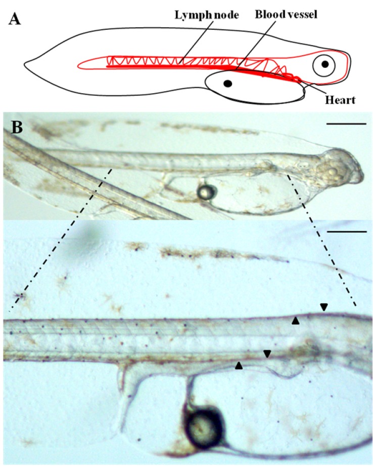
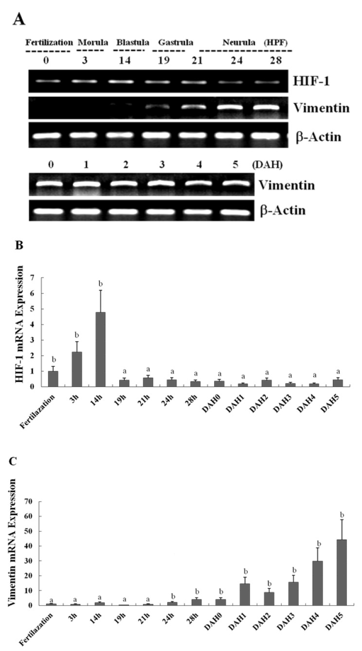
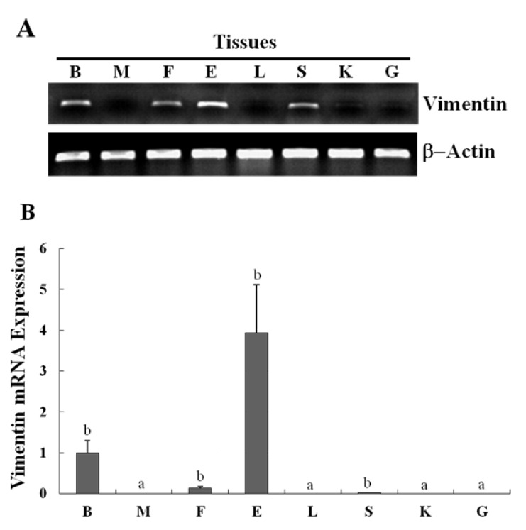
Cardiovascular system is the primary organ to develop and reach a functional state, which underscores the essential role of the vasculature in the developing embryo. The vasculature is a highly specialized organ that functions in a number of key physiological works including the carrying of oxygen and nutrients to tissues. It is closely involved in the formation of heart, and hence it is essential for survival during the hatching period. The expression of genes involved during vascular development in the olive flounder (Paralichthys olivaceus) in the days after hatching is not fully understood. Therefore, we examined the expression patterns of genes activated during the development of flounder. Microscopic observations showed that formation of blood vessels is related to the expression of the vimentin gene. Also, the temporal expression patterns of this vimentin-like gene in the developmental stages and in the normal tissues of olive flounder. The purpose of this study was to examine the expression patterns of vimentin in normal tissues of the olive flounder and during the development of the vascular system in newly hatched olive flounders and HIF-1 plays a vital role in the formation of blood vessels during development. Vimentin expression was strong at the beginning of the development of blood vessels, and was present throughout all developmental stages. Our findings have important implications with respect to the roles of vimentin and HIF-1 in the development and evolution of the first blood vessels in olive flounder. Further studies are required to elucidate the vimentin-mediated hypoxic response signal transduction and to decipher the functional role of vimentin in developmental stages.
Keywords: Intermediate Filament (IF); Olive flounder.; Vascular development; Vimentin.
Figures
References
-
- Chung AS, Ferrara N. Developmental and pathological angiogenesis. Annu Rev Cell Dev Biol. 2007;27:563–584. - PubMed
-
- Dunwoodie SL. The role of hypoxia in development of the mammalian embryo. Dev Cell. 2009;17:755–773. - PubMed
-
- Freeburg PB, Abrahamson DR. Hypoxia-inducible factors and kidney vascular development. J Am Soc Nephrol. 2003;14:2723–2730. - PubMed
-
- Fuchs E, Cleveland DW. A structural scaffolding of intermediate filaments in health and disease. Science. 1998;279:514–519. - PubMed
-
- Gariano RF, Sage EH, Kaplan HJ, Hendrickson AE. Development of astrocytes and their relation to blood vessels in fetal monkey retina. Invest Ophthalmol Vis Sci. 1996;37:2367–2375. - PubMed
LinkOut - more resources
Full Text Sources
Other Literature Sources
Miscellaneous