Effect of Long Term Reverse Feeding on the Reproductive and Non-reproductive Tissues in Male Mice
- PMID: 25949185
- PMCID: PMC4282209
- DOI: 10.12717/DR.2014.18.3.161
Effect of Long Term Reverse Feeding on the Reproductive and Non-reproductive Tissues in Male Mice
Abstract
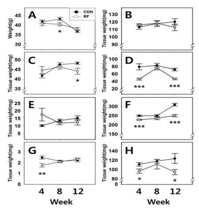
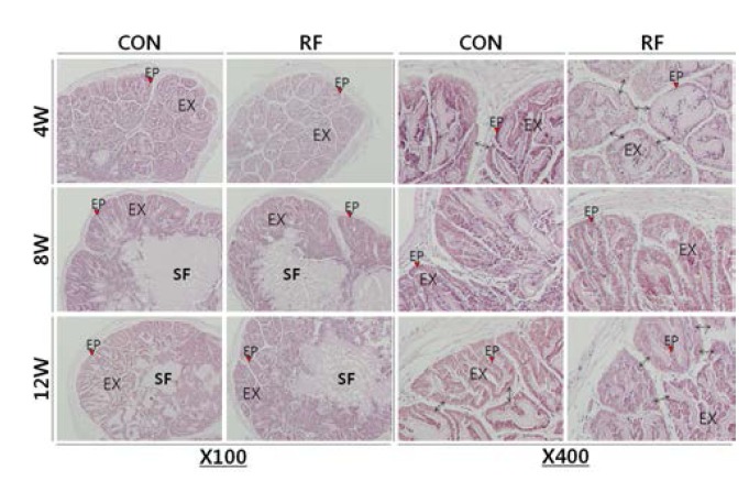
Previously, we demonstrated that the shift and/or restriction of feeding time during relatively short-term period (4 weeks) could alter the pituitary gonadotropin expression and the weights of seminal vesicle and prostate in rats. We also found that the reverse feeding (RF) schedule (up to 8 weeks) might induce an adaptable metabolic stress and cause impairment of androgen-dependent reproductive tissues. In the present study, we extended the RF time regimen up to 12 weeks, and measured the reproductive tissue weights. After 4 and 8 weeks of RF, the weights of epididymis were not significantly different. After 12 weeks, however, epididymis weights of RF animals were significantly different (CON 12W : RF 12W = 48.26±0.62 mg : 44.05±1.57 mg, p<0.05). After 4 and 12 weeks of feeding, seminal vesicle weights of RF animals were significantly decreased (CON 4W : RF 4W = 79.36±8.34 mg : 46.28±2.43 mg, p<0.001; CON 12W : RF 12W = 72.04±3.76 mg : 46.71±2.27 mg, p<0.001, respectively). Prostate weights were not changed by RF. Kidney and spleen weights of RF animals were significantly different on weeks 4 and 12 (Kidney, CON 4W : RF 4W = 249.72±4.20 mg : 228.41±3.03 mg, p<0.001; CON 12W : RF 12W = 309.15±7.49 mg : 250.72±6.13 mg, p<0.001, respectively, Spleen, CON 4W : RF 4W = 111.26±3.76 mg : 96.88±4.69 mg, p<0.05; CON 12W : RF 12W = 123.93±10.72 mg : 94.68±5.65 mg, p<0.05, respectively). Histology analysis of seminal vesicle revealed that the thinner epithelial cell layers, reduced complexities of swollen papilla folding in the exocrine glands on weeks 4 and 12 of RF. There was no histological difference between control and RF group on week 8. The present study indicates that up to 12 weeks RF induced differential changes in tissue weights of male mice. In particular, seminal vesicle, kidney and spleen seemed to temporarily adapted to the RF-induced metabolic stress on week 8 of feeding schedule. These results confirmed the our previous study that the RF might induce an adaptable metabolic stress and cause impairment of androgen-dependent reproductive tissues such as epididymis and seminal vesicle as well as non-reproductive tissues such as kidney and spleen. Further studies will be needed to achieve a better understanding of the how does mealtime shift affect the reproductive function and exact nature of adaptation.
Keywords: Circadian rhythm; Male mice; Reverse feeding (RF); Seminal vesicle; Tissue weight.
Figures
References
-
- Akiyama S, Ohta H, Watanabe S, Moriya T, Hariu A, Nakahata N, Chisaka H, Matsuda T, Kimura Y, Tsuchiya S, Tei H, Okamura K, Yaegashi N. The uterus sustains stable biological clock during pregnancy. Tohoku J Exp Med. 2010;221:287–298. - PubMed
-
- Alvarez JD, Chen D, Storer E, Sehgal A. Non-cyclic and developmental stage-specific expression of circadian clock proteins during murine spermatogenesis. Biol Reprod. 2003;69:81–91. - PubMed
-
- Boden MJ, Kennaway DJ. Circadian rhythms and reproduction. Reproduction. 2006;132:379–392. - PubMed
-
- Boden MJ, Varcoe TJ, Kennaway DJ. Circadian regulation of reproduction: from gamete to offspring. Prog Biophys Mol Biol. 2013;113:387–397. - PubMed
-
- Cagnacci A, Soldani R, Melis GB, Volpe A. Diurnal rhythms of labor and delivery in women: modulation by parity and seasons. Am J Obstet Gynecol. 1998;178(1 Pt 1):140–145. - PubMed
LinkOut - more resources
Full Text Sources
Other Literature Sources