. 2015:2015:619720.
doi: 10.1155/2015/619720. Epub 2015 Apr 8.

Ferulic Acid Attenuates TGF-β1-Induced Renal Cellular Fibrosis in NRK-52E Cells by Inhibiting Smad/ILK/Snail Pathway

Affiliations

Ferulic Acid Attenuates TGF-β1-Induced Renal Cellular Fibrosis in NRK-52E Cells by Inhibiting Smad/ILK/Snail Pathway

Ming-Gang Wei et al. Evid Based Complement Alternat Med. 2015.

Abstract

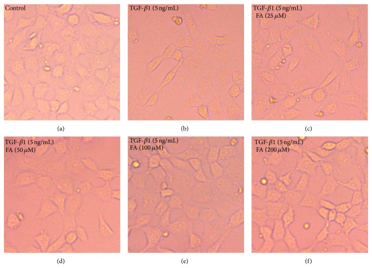
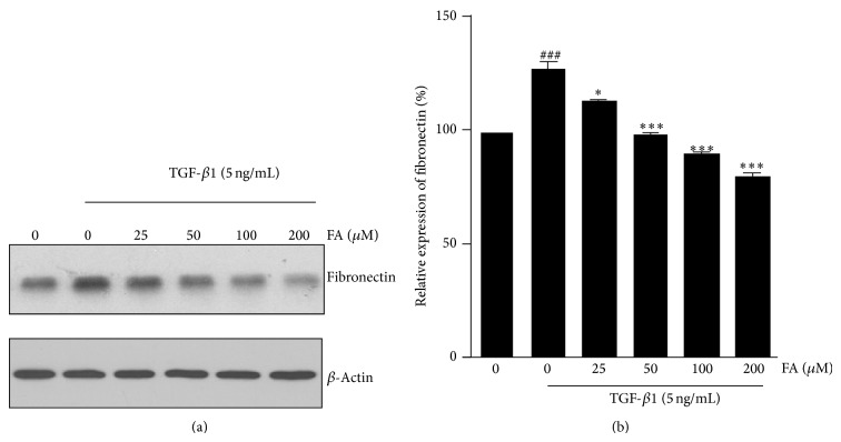
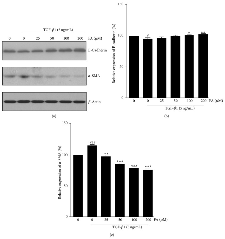
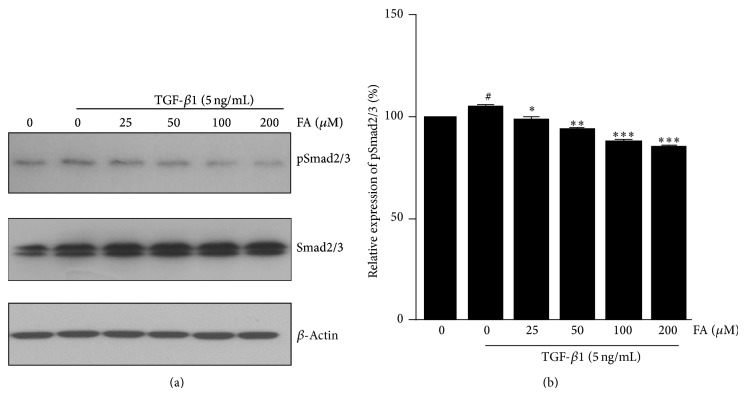
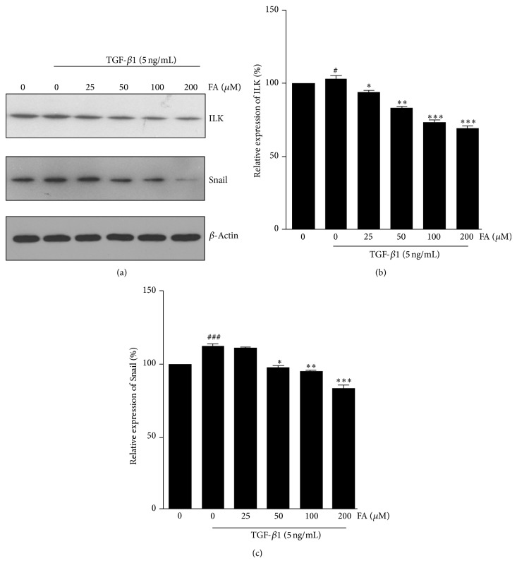
Renal fibrosis is a common cause of renal dysfunction with chronic kidney disease. Central to this process is epithelial-mesenchymal transformation (EMT) of proximal tubular epithelial cells driven by transforming growth factor-β1 (TGF-β1) signaling. The present study aimed to investigate the effect of Ferulic acid (FA) on EMT of renal proximal tubular epithelial cell line (NRK-52E) induced by TGF-β1 and to elucidate its underlying mechanism against EMT related to TGF-β1/Smads pathway. The NRK-52E cells were treated for 48 h with TGF-β1 (5 ng/mL) in different concentrations of FA (0 to 200 µM). Fibronectin, a mesenchymal marker, was assessed by western blotting. Western blotting was also used to examine the EMT markers (E-cadherin, and α-smooth muscle actin (α-SMA)), signal transducer (p-Smad2/3), and EMT initiator (Snail). ILK was also assayed by western blotting. The results showed that TGF-β1 induced spindle-like morphological transition in NRK-52E cells. Smad2/3 signaling pathway activation, increased fibronectin, α-SMA, ILK, and Snail expression, and decreased E-cadherin expression in TGF-β1-treated NRK-52E cells. FA efficiently blocked P-Smad2/3 activation and attenuated all these EMT changes induced by TGF-β1. These findings suggest that FA may serve as a potential fibrosis antagonist for renal proximal tubule cells by inhibiting EMT process.


Figures

Figure 1
Figure 1
FA reversed TGF-β1-induced morphological changes in NRK-52E cells. NRK-52E cells were incubated with 5 ng/mL of TGF-β1 for 48 h with different concentrations of FA (0, 25, 50, 100, and 200 μM). (a) Untreated NRK-52E cells showed that a pebble-like shape is clearly observed. (b) TGF-β1-treated cells showed a decrease in cell-cell contacts and adopt a more elongated morphological shape. (c), (d), (e), and (f) showed reversed TGF-β1-induced morphological changes by different concentrations of FA (magnification of 200x).
Figure 2
Figure 2
Effects of FA on TGF-β1-induced expression of fibronectin in NRK-52E cells. NRK-52E cells were incubated with 5 ng/mL of TGF-β1 for 48 h with different concentrations of FA (0, 25, 50, 100, and 200 μM). (a) The intercellular fibronectin was measured by western blotting with β-actin used as an internal control. (b) The fibronectin was quantitatively analyzed with Image J software. The data showing mean ± SD. # P < 0.05, ## P < 0.01, and ### P < 0.005 versus control (0 ng/mL TGF-β1). P < 0.05, ∗∗ P < 0.01, and ∗∗∗ P < 0.005 versus control (0 μM FA in the presence of 5 ng/mL TGF-β1).
Figure 3
Figure 3
Effects of FA on TGF-β1-induced expression of E-cadherin and α-SMA in NRK-52E cells. NRK-52E cells were incubated with 5 ng/mL of TGF-β1 for 48 h with different concentrations of FA (0, 25, 50, 100, and 200 μM). (a) Expression of E-cadherin and α-SMA at the protein level were determined by Western blotting with β-actin used as an internal control. (b) and (c) The expression level was quantitatively analyzed with Image J software. The data showing mean ± SD. # P < 0.05, ## P < 0.01, and ### P < 0.005 versus control (0 ng/mL TGF-β1). P < 0.05, ∗∗ P < 0.01, and ∗∗∗ P < 0.005 versus control (0 μM FA in the presence of 5 ng/mL TGF-β1).
Figure 4
Figure 4
Effects of FA on TGF-β1-induced expression of pSmad2/3 in NRK-52E cells. NRK-52E cells were incubated with 5 ng/mL of TGF-β1 for 48 h with different concentrations of FA (0, 25, 50, 100, and 200 μM). (a) Expression of Smad2/3 and pSmad2/3 at the protein level was determined by western blotting with β-actin used as an internal control. (b) The pSmad2/3 was quantitatively analyzed with Image J software. The data showing mean ± SD. # P < 0.05, ## P < 0.01, and ### P < 0.005 versus control (0 ng/mL TGF-β1). P < 0.05, ∗∗ P < 0.01, and ∗∗∗ P < 0.005 versus control (0 μM FA in the presence of 5 ng/mL TGF-β1).
Figure 5
Figure 5
Effects of FA on TGF-β1-induced expression of ILK and Snail in NRK-52E cells. NRK-52E cells were incubated with 5 ng/mL of TGF-β1 for 48 h with different concentrations of FA (0, 25, 50, 100, and 200 μM). (a) Expression of ILK and Snail at the protein level were determined by western blotting with β-actin used as an internal control. (b) and (c) The expression level was quantitatively analyzed with Image J software. The data showing mean ± SD. # P < 0.05, ## P < 0.01, and ### P < 0.005 versus control (0 ng/mL TGF-β1). P < 0.05, ∗∗ P < 0.01, and ∗∗∗ P < 0.005 versus control (0 μM FA in the presence of 5 ng/mL TGF-β1).

Similar articles

Cited by

References

    1. Boor P., Ostendorf T., Floege J. Renal fibrosis: novel insights into mechanisms and therapeutic targets. Nature Reviews Nephrology. 2010;6(11):643–656. doi: 10.1038/nrneph.2010.120. - DOI - PubMed
    1. Kriz W., Kaissling B., Le Hir M. Epithelial-mesenchymal transition (EMT) in kidney fibrosis: fact or fantasy? The Journal of Clinical Investigation. 2011;121(2):468–474. doi: 10.1172/jci44595. - DOI - PMC - PubMed
    1. Savagner P. Leaving the neighborhood: molecular mechanisms involved during epithelial-mesenchymal transition. BioEssays. 2001;23(10):912–923. doi: 10.1002/bies.1132. - DOI - PubMed
    1. Iwano M., Plieth D., Danoff T. M., Xue C., Okada H., Neilson E. G. Evidence that fibroblasts derive from epithelium during tissue fibrosis. The Journal of Clinical Investigation. 2002;110(3):341–350. doi: 10.1172/jci200215518. - DOI - PMC - PubMed
    1. Yang J., Liu Y. Dissection of key events in tubular epithelial to myofibroblast transition and its implications in renal interstitial fibrosis. The American Journal of Pathology. 2001;159(4):1465–1475. doi: 10.1016/s0002-9440(10)62533-3. - DOI - PMC - PubMed

LinkOut - more resources