

What proportions of focal liver lesions detected by unenhanced ultrasound are inconclusive?
- PMID: 25949268
- PMCID: PMC4412878
- DOI: 10.1177/1742271X14562995
What proportions of focal liver lesions detected by unenhanced ultrasound are inconclusive?
Abstract
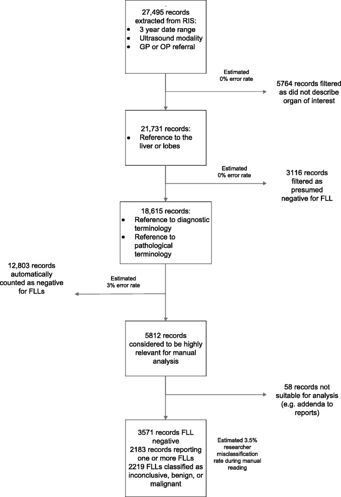
In August 2012, the National Institute for Health and Care Excellence produced positive diagnostics guidance on the ultrasound contrast agent SonoVue®, but recommended further research involving an estimation of the proportion of unenhanced ultrasound scans reporting, but not characterising, focal liver lesions, particularly in cirrhotic livers. Patient records from the Radiology Information System of an acute hospital trust were progressively filtered based on categorical fields and keywords in the free text reports, to obtain ultrasound records including the liver that were appropriate for manual analysis. In total, 21,731 records referred from general practice or out-patient clinics were analysed. Patients described as having cirrhosis were analysed as a subgroup. After automatic exclusion of records considered likely to be negative, 5812 records were manually read and categorised as focal liver lesion inconclusive, benign or malignant. In the general practice cohort of 9175 records, 746 reported the presence of one or more focal liver lesions, with 18.4% (95% CI 15.7% to 21.3%) of these records mentioning an inconclusive focal liver lesion. In the out-patient cohort of 12,556 records, 1437 reported one or more focal liver lesions, and 29.4% (95% CI 26.9% to 32.0%) of these were inconclusive. Cirrhosis was reported in 10.8% of the out-patient scans that also reported a focal liver lesion, and 47.4% (95% CI 39.3% to 55.6%) of these scans had an inconclusive focal liver lesion, compared with 27.3% (95% CI 24.9% to 29.8%) that were inconclusive in non-cirrhotic livers (odds ratio 2.4; 95% CI 1.7 to 3.4). This retrospective study indicates that unenhanced ultrasound scans, in which a focal liver lesion is detected, are frequently inconclusive, with the probability of an inconclusive scan being greater in out-patient than general practice referrals. Inconclusive focal liver lesions were also reported in greater proportions of cirrhotic than non-cirrhotic livers. The results of this research will inform future updates of National Institute for Health and Care Excellence diagnostics guidance.
Keywords: Liver; National Institute for Health and Care Excellence; SonoVue®; cirrhosis; inconclusive; ultrasound.
Figures
Similar articles
-
Heterogeneity in patient diagnostic pathways: an example from contrast-enhanced ultrasound diagnostic scans for focal liver lesions.BMC Res Notes. 2014 Mar 31;7:199. doi: 10.1186/1756-0500-7-199. BMC Res Notes. 2014. PMID: 24679189 Free PMC article.
-
Value of contrast-enhanced ultrasound (CEUS) in Focal Liver Lesions (FLL) with inconclusive findings on cross-sectional imaging.Clin Hemorheol Microcirc. 2020;74(3):327-339. doi: 10.3233/CH-190718. Clin Hemorheol Microcirc. 2020. PMID: 31658052
-
Double-contrast MRI (DC-MRI) in the study of the cirrhotic liver: utility of administering Gd-DTPA as a complement to examinations in which SPIO liver uptake and distribution alterations (SPIO-LUDA) are present and in the identification and characterisation of focal lesions.Radiol Med. 2006 Dec;111(8):1087-102. doi: 10.1007/s11547-006-0107-3. Epub 2006 Dec 20. Radiol Med. 2006. PMID: 17171525 English, Italian.
-
[Contrast ultrasound imaging in focal liver lesions: diagnostic value and guidelines].J Radiol. 2004 May;85(5 Pt 2):680-9. doi: 10.1016/s0221-0363(04)97649-4. J Radiol. 2004. PMID: 15238869 Review. French.
-
Focal masses in a non-cirrhotic liver: The additional benefit of CEUS over baseline imaging.Eur J Radiol. 2015 Sep;84(9):1636-43. doi: 10.1016/j.ejrad.2015.05.007. Epub 2015 May 22. Eur J Radiol. 2015. PMID: 26049958 Review.
Cited by
-
Multifocal Nodular Fatty Infiltration of the Liver: A Case Report of a Challenging Diagnostic Problem.Am J Case Rep. 2016 Mar 27;17:196-202. doi: 10.12659/ajcr.897283. Am J Case Rep. 2016. PMID: 27017525 Free PMC article.
-
Characterization of a hepatic haemangioma with contrast-enhanced ultrasound in an infant.Ultrasound. 2018 Aug;26(3):178-181. doi: 10.1177/1742271X17733298. Epub 2017 Oct 19. Ultrasound. 2018. PMID: 30147742 Free PMC article.
-
Toward the Development of Data Governance Standards for Using Clinical Free-Text Data in Health Research: Position Paper.J Med Internet Res. 2020 Jun 29;22(6):e16760. doi: 10.2196/16760. J Med Internet Res. 2020. PMID: 32597785 Free PMC article.
References
-
- Claudon M, Dietrich CF, Choi BI, et al. Guidelines and good clinical practice recommendations for contrast enhanced ultrasound (CEUS) in the liver–update 2012: a WFUMB-EFSUMB initiative in cooperation with representatives of AFSUMB, AIUM, ASUM, FLAUS and ICUS. Ultraschall in Med 2013; 34: 11–29. - PubMed
-
- National Institute for Health and Clinical Excellence. SonoVue (sulphur hexafluoride microbubbles) – contrast agent for contrast enhanced ultrasound imaging of the liver, London: NICE, 2012.
-
- Westwood M, Joore M, Grutters J, et al. Contrast-enhanced ultrasound using SonoVue (sulphur hexafluoride microbubbles) compared with contrast-enhanced computed tomography and contrast-enhanced magnetic resonance imaging for the characterisation of focal liver lesions and detection of liver metastases: a systematic review and cost-effectiveness analysis. Health Tech Assess 2013; 17: 1–243. - PMC - PubMed
-
- Thompson Coon J, Rogers G, Hewson P, et al. Surveillance of cirrhosis for hepatocellular carcinoma: systematic review and economic analysis. Health Tech Assess 2007; 11: 1–206. - PubMed
LinkOut - more resources
Full Text Sources
Other Literature Sources