. 2015 May 7:5:9788.
doi: 10.1038/srep09788.

Using "Tender" X-ray Ambient Pressure X-Ray Photoelectron Spectroscopy as A Direct Probe of Solid-Liquid Interface

Affiliations

Using "Tender" X-ray Ambient Pressure X-Ray Photoelectron Spectroscopy as A Direct Probe of Solid-Liquid Interface

Stephanus Axnanda et al. Sci Rep. .

Abstract

We report a new method to probe the solid-liquid interface through the use of a thin liquid layer on a solid surface. An ambient pressure XPS (AP-XPS) endstation that is capable of detecting high kinetic energy photoelectrons (7 keV) at a pressure up to 110 Torr has been constructed and commissioned. Additionally, we have deployed a "dip &pull" method to create a stable nanometers-thick aqueous electrolyte on platinum working electrode surface. Combining the newly constructed AP-XPS system, "dip &pull" approach, with a "tender" X-ray synchrotron source (2 keV-7 keV), we are able to access the interface between liquid and solid dense phases with photoelectrons and directly probe important phenomena occurring at the narrow solid-liquid interface region in an electrochemical system. Using this approach, we have performed electrochemical oxidation of the Pt electrode at an oxygen evolution reaction (OER) potential. Under this potential, we observe the formation of both Pt(2+) and Pt(4+) interfacial species on the Pt working electrode in situ. We believe this thin-film approach and the use of "tender" AP-XPS highlighted in this study is an innovative new approach to probe this key solid-liquid interface region of electrochemistry.
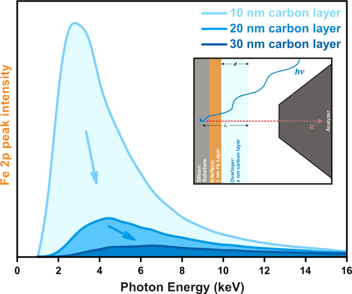

Figures

Figure 1
Figure 1
Integrated area of Fe 2p for a 1 nm Fe interface layer as a function of photon energy buried under various thicknesses of carbon to illustrate the ideal photon energy region for studying interface phenomena in systems on the order of 10’s of nm in thickness. The inset is a representation of the electrolyte, electrode, and electrolyte-electrode interface layer used in the SESSA simulation used to generate the data for this plot.
Figure 2
Figure 2
(a) Schematic top view of the “tender” X-ray AP-XPS analyzer and analysis chamber. (b) Detailed representation of the analysis chamber. (c) Photo of the actual “tender” X-Ray AP-XPS analyzer and the analysis chamber that is directly connected to the analyzer.
Figure 3
Figure 3
(a) Au 4f spectra of Au foil in Ar gas at different pressures, (b) integrated intensity of the Au 4f spectra relative to the vacuum condition as a function of Ar pressure, and (c) Au 4f of Au foil in Ar gas at pressure ranges from 50 Torr to 110 Torr using a 0.1 mm diameter aperture cone.
Figure 4
Figure 4
Schematic of three-electrode electrochemistry setup in the AP-XPS chamber. (a) Positions of electrodes before immersion and corresponding representative Pt 4f, O 1s spectra and electrochemical profile. (b) Electrodes are immersed in the electrolyte, where any electrochemical treatment can be performed within the AP-XPS chamber. Shown is a representative Pt foil CV in 6 M KF aqueous electrolyte. (c) Electrodes are placed at the AP-XPS measurement position, and corresponding representative Pt 4f and O 1s of the partially removed electrodes are overlaying the representative vapor exposed electrode spectra (shown in Fig. 4a). (d) is an image of the 3-electrode apparatus that has been “dip & pulled” from the electrolyte in the beaker and placed into XPS position while ensuring all three electrodes are in contact with the electrolyte within the beaker.
Figure 5
Figure 5
(a) Pt 4f of clean Pt foil in 1 × 10−3 Torr (grey), under 20 Torr of water vapor pressure (light blue), and under 0.8 V potential with CV treatment (dark blue), (b) integrated intensity of the Pt 4f spectra relative to the UHV condition at different treatment conditions.
Figure 6
Figure 6
(a) Pt 4f, (b) O 1s, and (c) K 2p of Pt foil after dipped in 6 M KF after cyclic voltammetry between −0.8 V and 0.8 V is applied, followed by holding the potential at −0.8 V and −0.4 V, and (d) Pt 4f with potential holding at 0 V and 1.2 V during the XPS measurement. All of the potentials are reported with respect to Ag/AgCl reference electrode.

References

    1. Hűfner S. Photoelectron Spectroscopy Principles and Applications. Springer 2003).
    1. Siegbahn K. ESCA applied to free molecules. North-Holland Pub. Co. 1969).
    1. Siegbahn H. Electron Spectroscopy for Chemical Analysis of Liquids and Solutions. J. Phys. Chem. B 89, 897 (1985).
    1. Frank Ogletree D., Bluhm H., Hebenstreit E. D. & Salmeron M. Photoelectron spectroscopy under ambient pressure and temperature conditions. Nucl. Instrum. Meth. A. 601, 151–160, doi: 10.1016/j.nima.2008.12.155 (2009). - DOI
    1. Salmeron M. & Schlögl R. Ambient pressure photoelectron spectroscopy: A new tool for surface science and nanotechnology. Surf. Sci. Rep. 63, 169–199 (2008).

Publication types