. 2015 May 7;6(5):e1748.
doi: 10.1038/cddis.2015.119.

The p38α mitogen-activated protein kinase is a key regulator of myelination and remyelination in the CNS

Affiliations

The p38α mitogen-activated protein kinase is a key regulator of myelination and remyelination in the CNS

S-H Chung et al. Cell Death Dis. .

Abstract

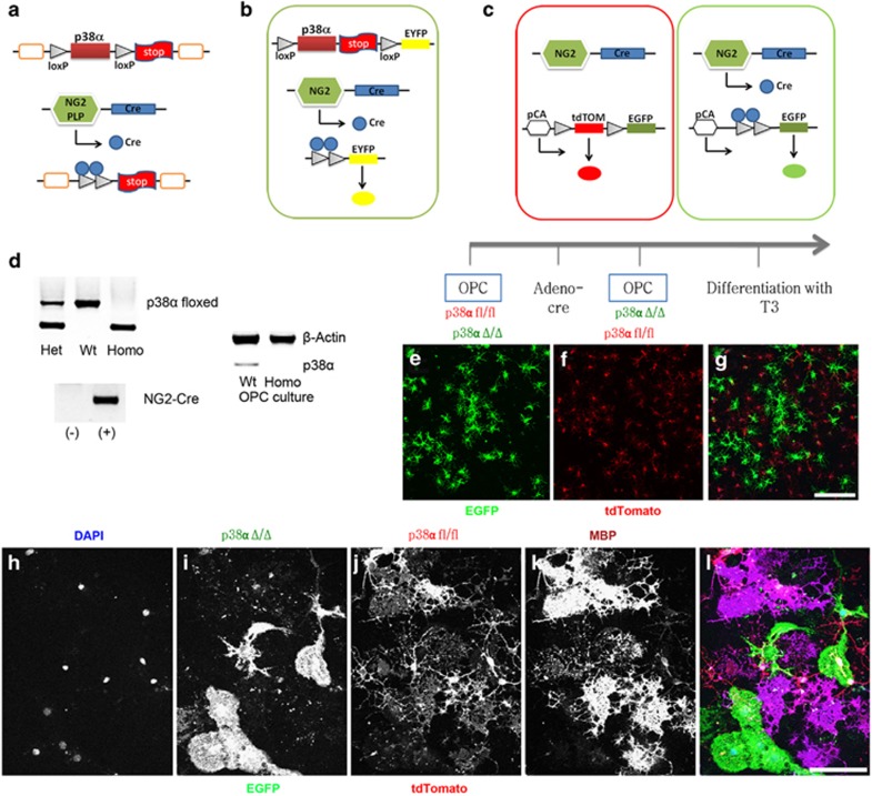
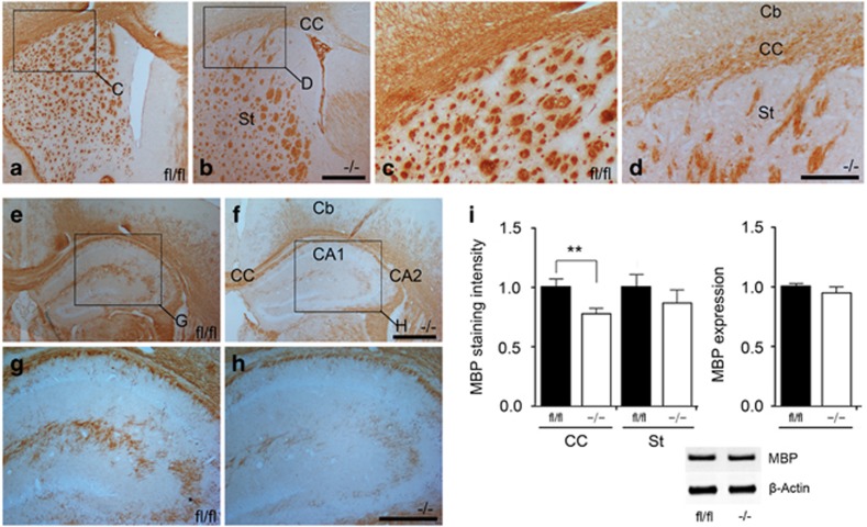
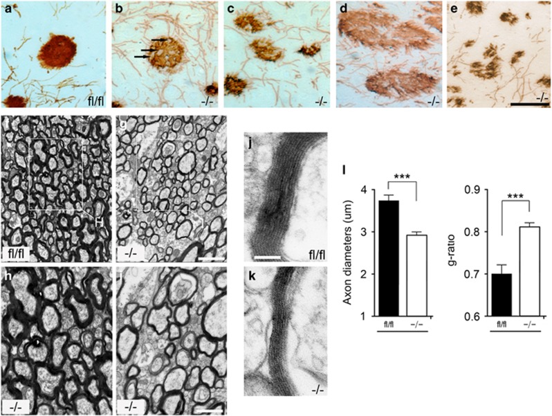
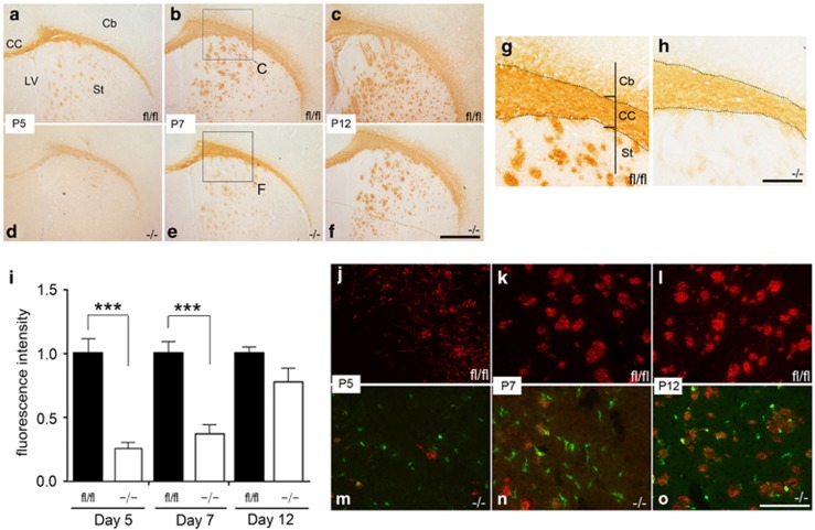
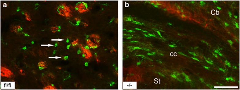
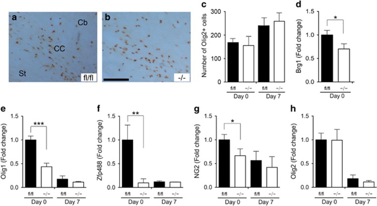
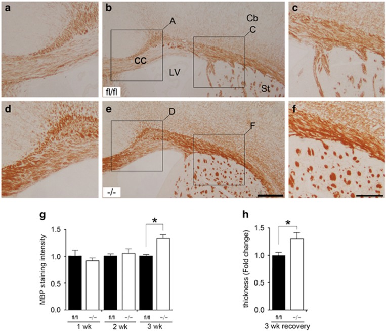
The p38α mitogen-activated protein kinase (MAPK) is one of the serine/threonine kinases regulating a variety of biological processes, including cell-type specification, differentiation and migration. Previous in vitro studies using pharmacological inhibitors suggested that p38 MAPK is essential for oligodendrocyte (OL) differentiation and myelination. To investigate the specific roles of p38α MAPK in OL development and myelination in vivo, we generated p38α conditional knockout (CKO) mice under the PLP and nerve/glial antigen 2 (NG2) gene promoters, as these genes are specifically expressed in OL progenitor cells (OPCs). Our data revealed that myelin synthesis was completely inhibited in OLs differentiated from primary OPC cultures derived from the NG2 Cre-p38α CKO mouse brains. Although an in vivo myelination defect was not obvious after gross examination of these mice, electron microscopic analysis showed that the ultrastructure of myelin bundles was severely impaired. Moreover, the onset of myelination in the corpus callosum was delayed in the knockout mice compared with p38α fl/fl control mice. A delay in OL differentiation in the central nervous system was observed with concomitant downregulation in the expression of OPC- and OL-specific genes such as Olig1 and Zfp488 during early postnatal development. OPC proliferation was not affected during this time. These data indicate that p38α is a positive regulator of OL differentiation and myelination. Unexpectedly, we observed an opposite effect of p38α on remyelination in the cuprizone-induced demyelination model. The p38α CKO mice exhibited better remyelination capability compared with p38α fl/fl mice following demyelination. The opposing roles of p38α in myelination and remyelination could be due to a strong anti-inflammatory effect of p38α or a dual reciprocal regulatory action of p38α on myelin formation during development and on remyelination after demyelination.


Figures

Figure 1
Figure 1
Generation of NG2/PlpCrep38α CKO mice and in vitro differentiation of p38α CKO OPCs. (a) Generation of p38α CKO mice by crossing NG2/Plp-Cre mice and mice with p38α-floxed alleles (p38α fl/fl). Schematic diagram showing the targeted genetic locus of p38α gene flanked by two loxP sites. p38α gene deletion is driven by Cre-loxP recombination that is under the control of NG2/Plp promoter activity in targeted cells. (b and c) Schematic illustrations demonstrating the EYFP (b) and ROSA26tdTomato (c) reporter system. Under the control of NG2/Plp promoter, Cre recombination results in an excision of tdTomato (mT) and the expression of EGFP (mG) in NG2/Plp-positive cells. (d) Genotyping of NG2-Cre mice demonstrating homozygosity of p38α-floxed allele and positive expression of NG2-Cre transgene. Western blotting analysis confirmed that the p38a protein is absent in p38α CKO OPC cultures. (eg) Schematic diagram of in vitro differentiation of purified primary OPCs from CKO mice. Four days after culture, both p38α CKO (green: e) and p38α fl/fl cells (red: f) were observed. (hl) Immunohistochemistry for anti-MBP after terminal differentiation of OPCs into myelin-forming OLs. MBP immunofluorescence labeling demonstrates that MBP expression was diminished in p38α CKO cells (green cells in panels (i) and (l)), while p38α fl/fl cells express MBP (red cells in panels (j) and (l)). Scale bars: panel (g)=500 μm; panel (l)=50 μm
Figure 2
Figure 2
No obvious gross myelination defects were observed in p38α CKO mice. (ad) Transverse sections were peroxidase stained for anti-MBP in p38α fl/fl (a and c) and p38α CKO (b and d) at P28. Panels (c) and (d) are higher magnification views of the regions indicated in panels (a) and (b). (eh) Transverse sections peroxidase stained for anti-MBP in p38α fl/fl (e and g) and p38α CKO (f and h) at P28. Panels (g) and (h) are higher magnification views of the regions indicated in panels (e) and (f). MBP expression in some areas of the CC seemed to be relatively weak in the knockout compared with p38α fl/fl, but generally the difference was not obvious at low magnification of light microscope level. (i) Statistical analysis of the MBP staining intensity in the CC (P<0.01) and St of P28 p38α CKO and p38α fl/fl mice. Western blotting analysis of MBP protein in the whole brains of P28 p38α CKO and p38α fl/fl mice. **P<0.01. Abbreviation: Cb, cerebral cortex. Scale bars: panel (b)=500 μm; panel (d)=250 μm; panel (f)=500 μm; panel (h)=250 μm
Figure 3
Figure 3
p38α CKO mice show morphological defects and reduced axon diameters. (ae) Sagittal sections were peroxidase stained for anti-MBP in p38α fl/fl (a) and p38α CKO (bd) at P12. Axonopathies observed in the p38α CKO: axonal swelling (b and c), changes in the density of axons (c and d) and degeneration (e) were frequently seen. (fk) Electron microscopic analysis of myelinated bundles in CC sections from P12 p38α CKO and p38α fl/fl mice. Panels (h) and (i) are higher magnification views of the regions indicated in panels (f) and (g). The thickness of myelin bundle diameter surrounding the axons was significantly reduced in the p38α CKO (k) compared with p38α fl/fl (j). (h) and (i) are higher magnification views of the regions indicated in panels (f) and (g). (l) Quantification of axon diameters and g-ratio (the ratio of axon diameter to fiber diameter). The axon diameters were significantly reduced in the CKO CC (P<0.0001). The g-ratio was significantly increased from 0.693 in p38α fl/fl to 0.818 in p38α CKO mice (P<0.0001). ***P<0.005. Scale bars: panel (e)=50 μm; panel (g)=5 μm; panel (h)=2.5 μm; panel (j)=1 μm
Figure 4
Figure 4
Delay in the onset of myelination in the CC of p38α CKO mice. (ah) Transverse sections peroxidase stained for anti-MBP at P5, P7 and P12 in p38α fl/fl and p38α CKO revealed that the onset and progression of myelination were delayed in the p38α CKO. Panels (g) and (h) are higher magnification views of the regions indicated in panels (b) and (e). (i) Quantitative analysis of MBP expression during different postnatal periods in the CC of the p38α fl/fl and p38α CKO mice, suggesting a delay in onset of myelination (P5 and P7 P<0.0001). (jo) Immunofluorescence-stained sections with anti-EYFP (green) and MBP (red) showed that the onset and progression of myelination were delayed in the p38α CKO CC. Green cells represent p38α knockout cells. Abbreviations: Cb, cerebral cortex; LV, lateral ventricle. ***P<0.005. Scale bars: panel (f)=250 μm; panel (h)=75 μm; panel (o)=250 μm
Figure 5
Figure 5
The p38α knockout cells show myelination defects in vivo. Immunofluorescence-stained sections with anti-EYFP (green) and MBP (red) in the (a) St and (b) CC of p38α CKO mice. Green cells represent p38α knockout cells. Majority of p38α knockout cells (arrows) did not express MBP. Abbreviation: Cb, cerebral cortex. Scale bar: panel (b)=100 μm
Figure 6
Figure 6
p38α gene deletion results in a delayed OL differentiation without affecting proliferation. (a and b) Transverse sections were peroxidase stained for anti-Olig2 at P7 in the CC of the p38α fl/fl (a) and p38α CKO mice (b). (c) Quantification of Olig2-immunopositive cells in the CC at P0 and P3 in the p38α fl/fl and p38α CKO. The numbers of Olig2-posivtive cells were unchanged. (dh) Quantitative PCR analysis for Brg1 (d), Olig1 (e), Zfp488 (f), NG2 (g) and olig2 (h) at P0 and P7 in the p38α fl/fl and p38α CKO mice. Olig1 and Zfp488 expression is significantly downregulated in the P0 p38α CKO mice (e and f). *P<0.05, **P<0.01 ***P<0.005. Abbreviation: Cb, cerebral cortex. Scale bar: panel (b)=100 μm
Figure 7
Figure 7
p38α CKO promotes remyelination after cuprizone-induced demyelination. (af) Transverse sections peroxidase stained with anti-MBP showing remyelination process in the Cc after 3 weeks following discontinuation of cuprizone intake in the p38α fl/fl (ac) and p38α CKO (df) mice. Panels (a and c) and (d and f) are higher magnification views of the regions indicated in panels (b) and (e), respectively. MBP expression is significantly increased in the CC of p38α CKO (df) compared with p38α fl/fl (ac) mice. (g and h) Quantification of MBP staining intensity (g) and thickness (h) during a recovery period in the p38α fl/fl and p38α CKO. p38α CKO mice showed a better remyelination ability compared with p38α fl/fl during the remyelination process. *P<0.05. Abbreviations: Cb, cerebral cortex; LV, lateral ventricle. Scale bar: panel (e)=500 μm; panel (f)=250 μm

References

    1. Gold R, Linington C, Lassmann H. Understanding pathogenesis and therapy of multiple sclerosis via animal models. 70 years of merits and culprits in experimental autoimmune encephalomyelitis research. Brain 2006; 129: 1953–1971. - PubMed
    1. Khwaja O, Volpe JJ. Pathogenesis of cerebral white matter injury of prematurity. Arch Dis Child Fetal Neonatal Ed 2008; 93: 153–161. - PMC - PubMed
    1. Deng W. Neurobiology of injury to the developing brain. Nat Rev Neurol 2010; 6: 328–336. - PubMed
    1. Baumann N, Pham-Dinh D. Biology of oligodendrocyte and myelin in the mammalian central nervous system. Physiol Rev 2001; 81: 871–927. - PubMed
    1. Miller RH. Regulation of oligodendrocyte development in the vertebrate CNS. Prog Neurobiol 2002; 67: 451–467. - PubMed

Publication types

Substances