. 2015 Jul;88(1051):20150157.
doi: 10.1259/bjr.20150157. Epub 2015 May 7.

Fatty lesions in and around the heart: a pictorial review

Affiliations

Fatty lesions in and around the heart: a pictorial review

R Pruente et al. Br J Radiol. 2015 Jul.

Abstract

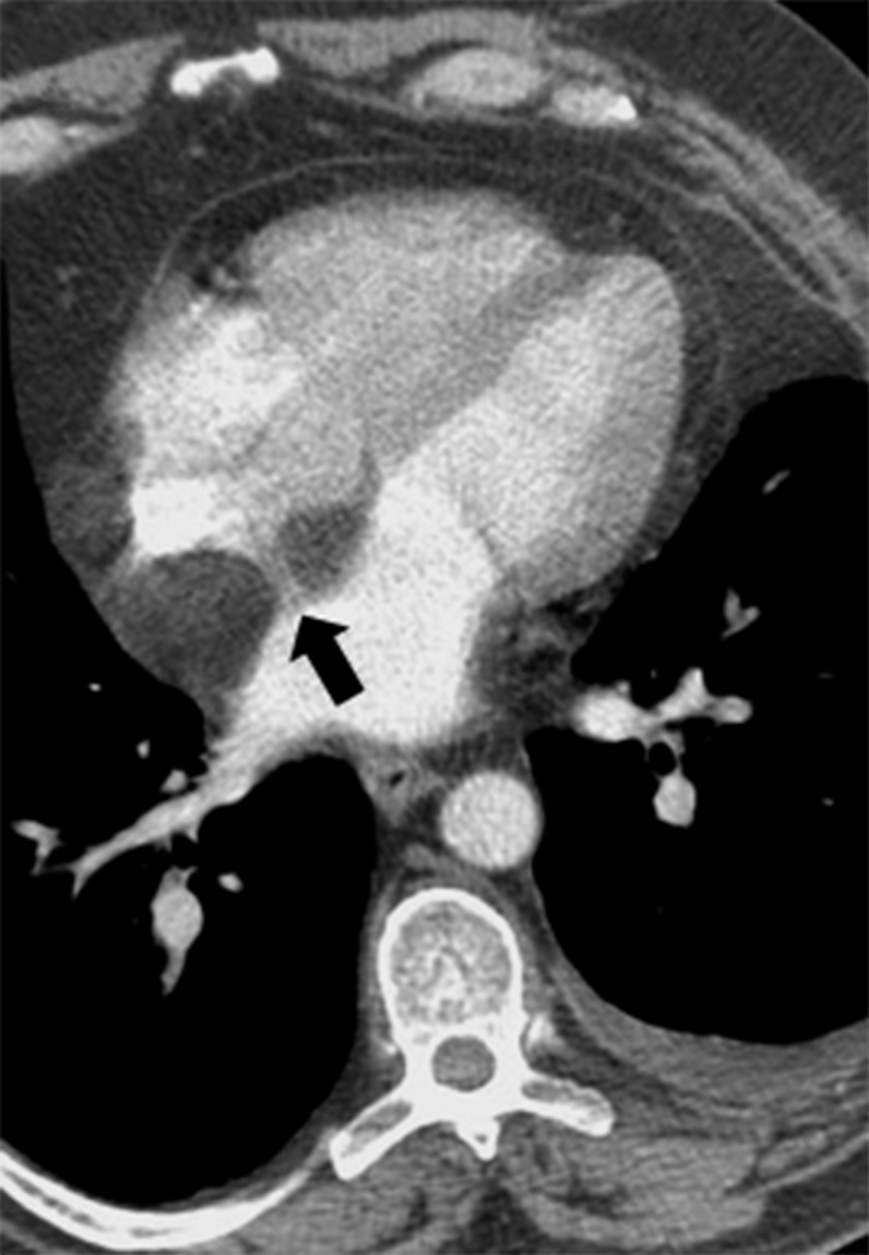
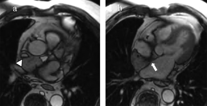
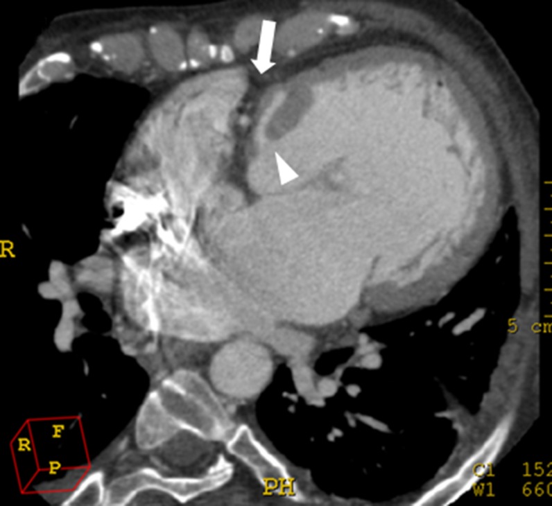
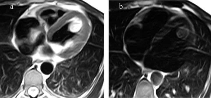
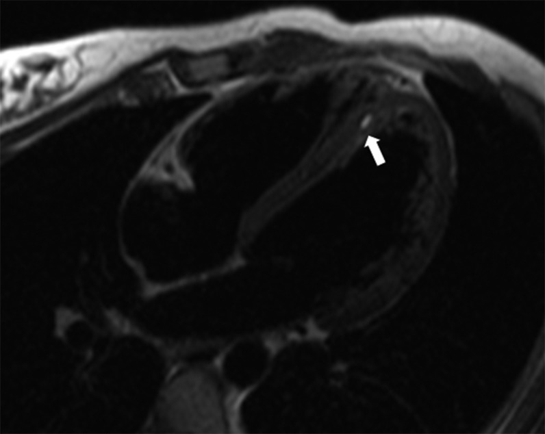
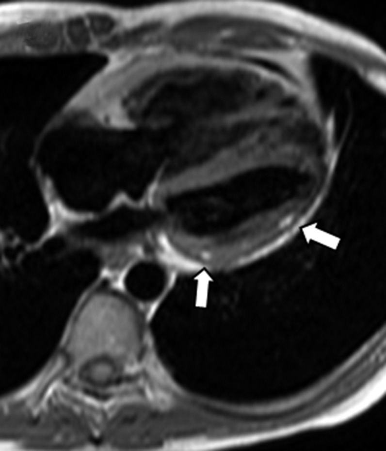
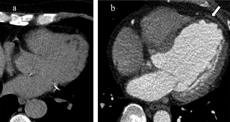
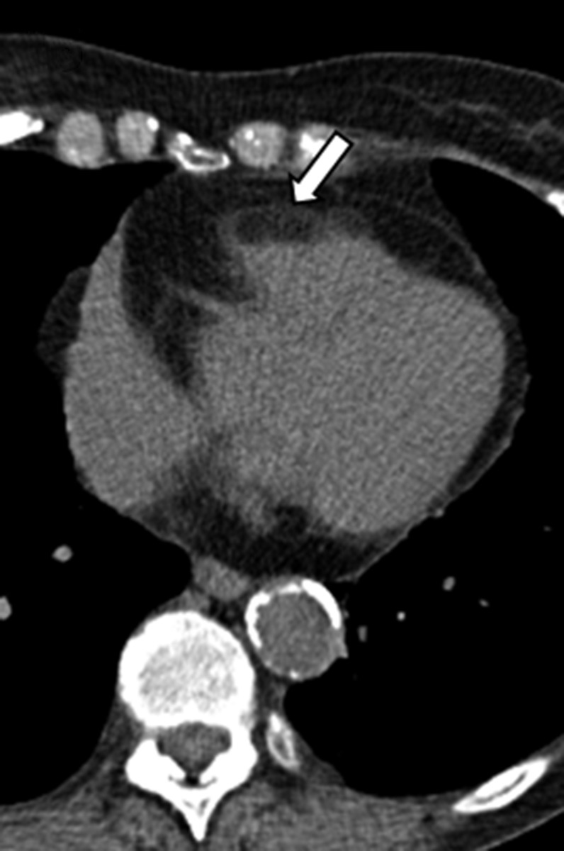
A wide variety of fat-containing entities occur in and near the heart. These findings are often encountered by radiologists and may be incidental or the reason for the patient's clinical presentation. Cross-sectional imaging helps to characterize the extent of these lesions and to formulate a differential diagnosis, which varies by lesion location, imaging features and patient demographics. The purpose of this pictorial essay is to familiarize radiologists with these fat-containing lesions and to help avoid misdiagnosis and errors in management. This pictorial review will discuss the normal fatty structures in and around the heart. A range of common and uncommon fat-containing lesions will then be reviewed based upon lesion location.


Figures

Figure 1.
Figure 1.
Flow chart providing an overview fat-containing lesions encountered in and near the heart, organized by location. ARVD, arrhythmogenic right ventricular dysplasia; LHIAS, lipomatous hypertrophy of the interatrial septum; MI, myocardial infract; RVOT, right ventricular outflow tract; TSC, tuberous sclerosis complex.
Figure 2.
Figure 2.
Mediastinal teratoma. Coronal contrast-enhanced CT image demonstrates a large, encapsulated mass in the right paracardiac region resulting in mass effect upon the right atrium and right superior pulmonary vein. Attenuation characteristics of this mass are consistent with a mature teratoma with foci of coarse calcification and a significant amount of fatty tissue.
Figure 3.
Figure 3.
Thymoliposarcoma. Transaxial contrast-enhanced CT image of the chest reveals a very large, predominantly fatty mass throughout the anterior mediastinum and extending along the cardiophrenic angles. There are several enhancing septa (arrow) as well as solid components. The mass results in posterior displacement of the mediastinal structures.
Figure 4.
Figure 4.
Epipericardial fat necrosis. Transaxial contrast-enhanced CT images of the chest demonstrate a lobulated low attenuation mass in the right anterior pericardiophrenic angle with intrinsic inflammatory stranding (arrows) consistent with epipericardial fat necrosis.
Figure 5.
Figure 5.
Fat-containing Morgagni hernia. Axial contrast-enhanced CT image shows a large amount of omental fat herniating through an anterior diaphragmatic defect along with loops of both small and large bowel. Reformatted images (not shown) depicted mesenteric vessels herniating through the defect as well.
Figure 6.
Figure 6.
Pericardial lipoma. Transaxial contrast-enhanced CT images at the level of the heart reveal a large low attenuating mass confined within the pericardium consistent with pericardial lipoma. Note how the lesion insinuates throughout the space it occupies. Also note the significant mass effect upon the superior vena cava (arrowhead) and pulmonary veins (arrow).
Figure 7.
Figure 7.
(a, b) Pericardial lipoblastoma in a young child. Coronal (a) and sagittal (b) T1 weighted images show a large fatty pericardial mass occupying the left hemithorax. Note the characteristic intratumoral streaks consistent with the internal septa (arrows). The imaging appearance appearance as well as the patient's age at presentation favoured a diagnosis of lipoblastoma over liposarcoma. Reproduced from Restrepo et al.
Figure 8.
Figure 8.
Epicardial lipomatosis. Axial contrast image of the chest demonstrates prominent epicardial (arrow) and mediastinal fat (arrowhead).
Figure 9.
Figure 9.
Lipomatous hypertrophy of the interatrial septum. Contrast-enhanced transaxial CT image of the heart shows invaginated fatty tissue in the interatrial septum and mildly encasing the superior vena cava. Note how the fossa ovalis is characteristically spared (arrow).
Figure 10.
Figure 10.
(a, b) Lipomatous hypertrophy of the interatrial septum in a 63-year-old male with history of right atrial mass seen on echocardiography. Balanced steady-state free precession MR images demonstrate a large amount of tissue in the interatrial septum. This tissue follows signal characteristics of fat on all sequences with signal loss on fat saturated sequences (not shown). Note narrowing of the superior vena cava (arrowhead) and sparing of the fossa ovalis (arrow).
Figure 11.
Figure 11.
Tricuspid atresia. Horizontal long axis reformat from a contrast-enhanced CT of the heart demonstrating a deep fatty bar in the region of the right atrioventricular groove and associated absence of tricuspid valvular tissue (arrow). As a result, the right ventricle is diminutive. A small ventricular septal defect is present (arrowhead).
Figure 12.
Figure 12.
(a, b) Cardiac lipoma. Transaxial T2 weighted fast turbo spin echo MR image (a) shows an encapsulated, hyperintense mass along the left ventricular wall. Fat saturated T1 weighted image (b) confirms the fatty content of the mass consistent with an intracardiac lipoma.
Figure 13.
Figure 13.
Intramyocardial fat deposits in tuberous sclerosis complex. Transaxial T2 weighted dark blood image in a patient with history of tuberous sclerosis reveals a small intramyocardial focus of fat (arrow).
Figure 14.
Figure 14.
Arrhythmogenic right ventricular dysplasia. Horizontal long axis T1 weighted image shows foci of intramyocardial high signal in the lateral wall of the left ventricle (arrows). Fat-suppressed images revealed nulling of signal in these foci with diffuse delayed enhancement of the right ventricle (not shown) characteristic of arrhythmogenic right ventricular dysplasia.
Figure 15.
Figure 15.
(a, b) Lipomatous metaplasia in chronic myocardial infarct in two different patients. Transaxial non-contrast CT image of the heart demonstrates curvilinear low attenuation in a subendocardial distribution along the anterolateral and apical walls of the left ventricle. In a different patient, there is transmural fibrofatty replacement and thinning along the interventricular septum, anteroseptal wall and apex of the left ventricle (arrow) consistent with prior myocardial infarct in the left anterior descending distribution.
Figure 16.
Figure 16.
Senescent fatty infiltration of the right ventricular outflow tract. Transaxial non-contrast CT image of the heart in an 81-year-old asymptomatic female shows extensive fatty infiltration of the right ventricular free wall extending into the right ventricular outflow tract (arrow).

References

    1. Gaerte SC, Meyer CA, Winer-Muram HT, Tarver RD, Conces DJ, Jr. Fat-containing lesions of the chest. Radiographics 2002; 22: S61–78. doi: 10.1148/radiographics.22.suppl_1.g02oc08s61 - DOI - PubMed
    1. Lamba G, Frishman WH. Cardiac and pericardial tumors. Cardiol Rev 2012; 20: 237–52. doi: 10.1097/CRD.0b013e31825603e7 - DOI - PubMed
    1. Molinari F, Bankier AA, Eisenberg RL. Fat-containing lesions in adult thoracic imaging. AJR Am J Roentgenol 2011; 197: W795–813. doi: 10.2214/AJR.11.6932 - DOI - PubMed
    1. Pineda V, Andreu J, Cáceres J, Merino X, Varona D, Domínguez-Oronoz R. Lesions of the cardiophrenic space: findings at cross-sectional imaging. Radiographics 2007; 27: 19–32. doi: 10.1148/rg.271065089 - DOI - PubMed
    1. Restrepo CS, Vargas D, Ocazionez D, Martínez-Jiménez S, Betancourt Cuellar SL, Gutierrez FR. Primary pericardial tumors. Radiographics 2013; 33: 1613–30. doi: 10.1148/rg.336135512 - DOI - PubMed