

Review
doi: 10.1371/journal.ppat.1004745.
eCollection 2015 May.
The elusive role of the prion protein and the mechanism of toxicity in prion disease
Affiliations
- PMID: 25951168
- PMCID: PMC4423772
- DOI: 10.1371/journal.ppat.1004745
Item in Clipboard
Review
The elusive role of the prion protein and the mechanism of toxicity in prion disease
PLoS Pathog.
.
No abstract available
Conflict of interest statement
The author has declared that no competing interests exist.
Figures

(A) PrPC consists of a flexible N terminus (mauve) and a globular C-terminal domain (green) attached to the plasma membrane (PM) by a GPI anchor (black line). PrPC associates with NMDARs, attenuating their activity [5]. (B–C) Interaction with extracellular PrPSc causes the N terminus of PrPC to undergo a structural rearrangement. This leads to aberrant interaction of PrPC with NMDARs and their hyperactivation (B) and/or abnormal insertion of the PrPC N terminus into the lipid bilayer with generation of a toxic pore (C). In addition to NMDARs, PrPC misfolding at the cell surface may corrupt the activity of other PrPC-interacting ion channels or signaling complexes.
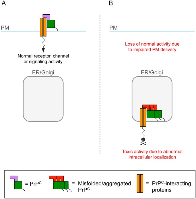
(A) PrPC on the plasma membrane (PM) influences the activity of neurotransmitter receptors, ion channels, and signaling complexes with which it interacts. (B) Owing to retention in transport organelles (ER/Golgi), misfolded/aggregated PrPC sequesters the interacting protein in intracellular compartments, leading to loss of normal function on the cell membrane [6]. Intracellular retention might also cause the complex to function abnormally and generate a toxic signal.
References
Publication types
MeSH terms
Substances
Grants and funding
LinkOut - more resources
Full Text Sources
Other Literature Sources
