Review
. 2016 Jan:40:1-23.
doi: 10.1016/j.yfrne.2015.04.003. Epub 2015 May 4.

Vasopressin and oxytocin receptor systems in the brain: Sex differences and sex-specific regulation of social behavior

Affiliations
Review

Vasopressin and oxytocin receptor systems in the brain: Sex differences and sex-specific regulation of social behavior

Kelly M Dumais et al. Front Neuroendocrinol. 2016 Jan.

Abstract

The neuropeptides vasopressin (VP) and oxytocin (OT) and their receptors in the brain are involved in the regulation of various social behaviors and have emerged as drug targets for the treatment of social dysfunction in several sex-biased neuropsychiatric disorders. Sex differences in the VP and OT systems may therefore be implicated in sex-specific regulation of healthy as well as impaired social behaviors. We begin this review by highlighting the sex differences, or lack of sex differences, in VP and OT synthesis in the brain. We then discuss the evidence showing the presence or absence of sex differences in VP and OT receptors in rodents and humans, as well as showing new data of sexually dimorphic V1a receptor binding in the rat brain. Importantly, we find that there is lack of comprehensive analysis of sex differences in these systems in common laboratory species, and we find that, when sex differences are present, they are highly brain region- and species-specific. Interestingly, VP system parameters (VP and V1aR) are typically higher in males, while sex differences in the OT system are not always in the same direction, often showing higher OT expression in females, but higher OT receptor expression in males. Furthermore, VP and OT receptor systems show distinct and largely non-overlapping expression in the rodent brain, which may cause these receptors to have either complementary or opposing functional roles in the sex-specific regulation of social behavior. Though still in need of further research, we close by discussing how manipulations of the VP and OT systems have given important insights into the involvement of these neuropeptide systems in the sex-specific regulation of social behavior in rodents and humans.

Keywords: Females; Humans; Males; OT receptor; Oxytocin; Rodents; Sex differences; Social behavior; V1a receptor; Vasopressin.


Figures

Fig. 1
Fig. 1
Representative coronal sections showing V1aR binding densities in forebrain areas of male (M) and female (F) Wistar rats. Receptor autoradiography was performed on 16 μm cryocut coronal brain sections according to Dumais et al. (2013) using the 125I linear VP antagonist [125I]-d(CH2)5(Tyr[Me])-AVP (Perkin Elmer, Shelton, CT) as tracer. Brain sections were exposed to film for 7 days. The optical density of V1aR binding was measured using Image J (NIH, http://rsb.info.nih.gov/ij/). Each measurement was subtracted by tissue background and V1aR binding densities were calculated by taking the mean of 4–10 (depending on the region being analyzed) bilateral measurements of the region of interest per rat. The data was converted to dpm/mg (disintegrations per minute/milligram tissue) using a [125I] standard microscale (American Radiolabeled Chemicals Inc., St Louis, MO). Compared to females, males have higher V1aR binding densities in the somatosensory cortex (SSC), piriform cortex (PC), nucleus of the lateral olfactory tract (LOT), medial posterior BNST (BNSTmp), anteroventral thalamus (AVthal), tuberal lateral hypothalamus (TuLH), stigmoid hypothalamus (StigH), and dentate gyrus (DG). No sex differences were found in the Islands of Calleja (ICj), nucleus accumbens (NAc), dorsal lateral septum (LSd), intermediate LS (LSi), ventral LS (LSv), lateral dorsal BNST (BNSTld), lateral posterior BNST (BNSTlp), lateral hypothalamus (LH), arcuate nucleus of the hypothalamus (ArcN), suprachiasmatic nucleus of the hypothalamus (SCN), interstitial nucleus of the posterior limb of the anterior commissure (IPAC), ventromedial thalamus (VMthal), and medial part of the CeA (CeAm). Regions which show sex differences in V1aR binding density are underlined.
Fig. 2
Fig. 2
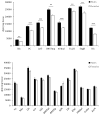
V1aR binding densities in forebrain regions of male and female Wistar rats. Males have higher V1aR binding densities than females in 8 out of 21 forebrain regions analyzed (top graph). These regions include the somatosensory cortex (SSC), piriform cortex (PC), nucleus of the lateral olfactory tract (LOT), medial posterior BNST (BNSTmp), anteroventral thalamus (AVthal), tuberal LH (TuLH), stigmoid hypothalamus (StigH), and dentate gyrus (DG). There are no sex differences in 13 forebrain regions analyzed (bottom graph). These regions include the Islands of Calleja (ICj), nucleus accumbens (NAc), dorsal lateral septum (LSd), intermediate LS (LSi), ventral LS (LSv), lateral dorsal BNST (BNSTld), lateral posterior BNST (BNSTlp), lateral hypothalamus (LH), arcuate nucleus of the hypothalamus (ArcN), suprachiasmatic nucleus of the hypothalamus (SCN), interstitial nucleus of the posterior limb of the anterior commissure (IPAC), ventromedial thalamus (VMthal), and medial part of the CeA (CeAm). Bars indicate means + SEM. **p<0.01, ***p<0.001, one-way ANOVA, correcting for multiple comparisons (FDR α≤0.0214)
Fig. 3
Fig. 3
Overlay of V1aR binding densities (blue) and OTR binding densities (red) in forebrain areas of male Wistar rats from adjacent coronal sections. The right column depicts representative rat brain images adapted from The Rat Brain Atlas (Paxinos & Watson, 1998). Overall, there is little overlap between V1aR binding and OTR binding profiles. Dense V1aR binding is found in the somatosensory cortex (SSc), piriform cortex (PC), Islands of Calleja (ICj), nucleus accumbens (NAc), lateral septum (LS), lateral dorsal BNST (not shown), lateral posterior BNST (BNSTlp), medial posterior BNST (not shown), nucleus of the lateral olfactory tract (LOT), dentate gyrus (DG), tuberal lateral hypothalamus (TuLH), anteroventral thalamus (AVthal), suprachiasmatic nucleus of the hypothalamus (not shown), interstitial nucleus of the posterior limb of the anterior commissure (IPAC), arcuate nucleus of the hypothalamus (ArcN), ventromedial thalamus (VMthal), and medial central amygdala (CeAm). Dense staining of OTR binding is found in the dorsal caudate putamen (CPu), agranular insular cortex (AIP), posterior BNST (BNSTp), medial preoptic area (MPOA), ventral medial hypothalamus (VMH), and lateral and capsular central amygdala (CeAl/c). Regions which show sex differences in V1aR or OTR binding are underlined.

References

    1. Albers HE. Species, sex and individual differences in the vasotocin/vasopressin system: Relationship to neurochemical signaling in the social behavior neural network. Front Neuroendocrinol 2014 - PMC - PubMed
    1. Albers HE, Rowland CM, Ferris CF. Arginine-vasopressin immunoreactivity is not altered by photoperiod or gonadal hormones in the Syrian hamster (Mesocricetus auratus) Brain Research. 1991;539(1):137–42. - PubMed
    1. Altemus M, Jacobson KR, Debellis M, Kling M, Pigott T, Murphy DL, Gold PW. Normal CSF oxytocin and NPY levels in OCD. Biol Psychiatry. 1999;45(7):931–3. - PubMed
    1. Armstrong WE. Hypothalamic supraoptic and paraventricular nuclei. In: Paxinos G, editor. The Rat Nervous System. San Diego, CA: Elsevier Academic; 2004. pp. 369–388.
    1. Asplund R, Aberg H. Diurnal variation in the levels of antidiuretic hormone in the elderly. Journal of Internal Medicine. 1991;229:131–4. - PubMed

Publication types

LinkOut - more resources