. 2015 May 8:6:7005.
doi: 10.1038/ncomms8005.

Topological control of the Caulobacter cell cycle circuitry by a polarized single-domain PAS protein

Affiliations

Topological control of the Caulobacter cell cycle circuitry by a polarized single-domain PAS protein

Stefano Sanselicio et al. Nat Commun. .

Abstract

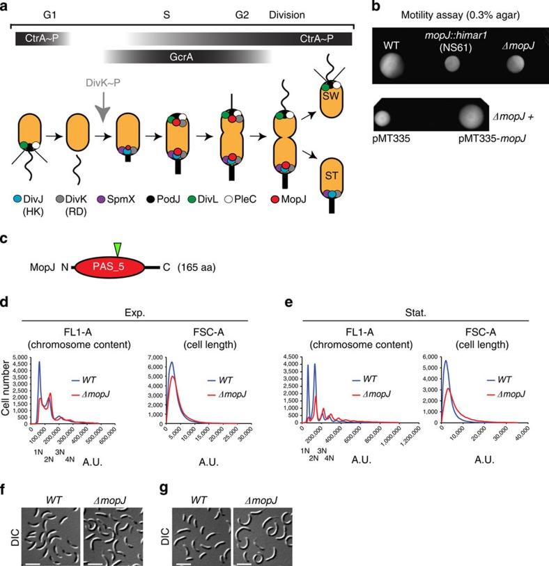
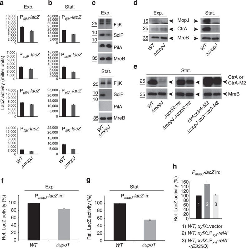
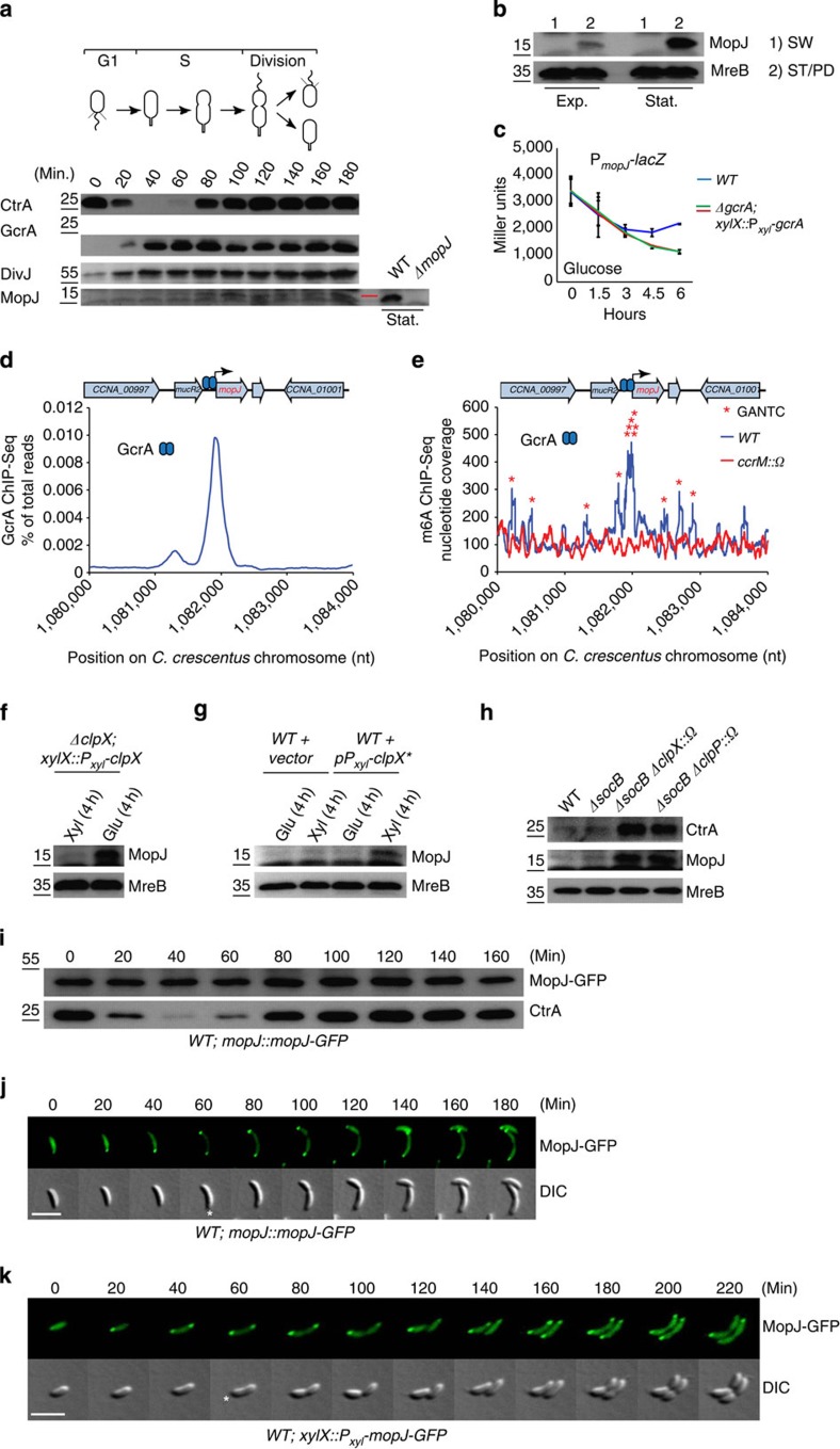
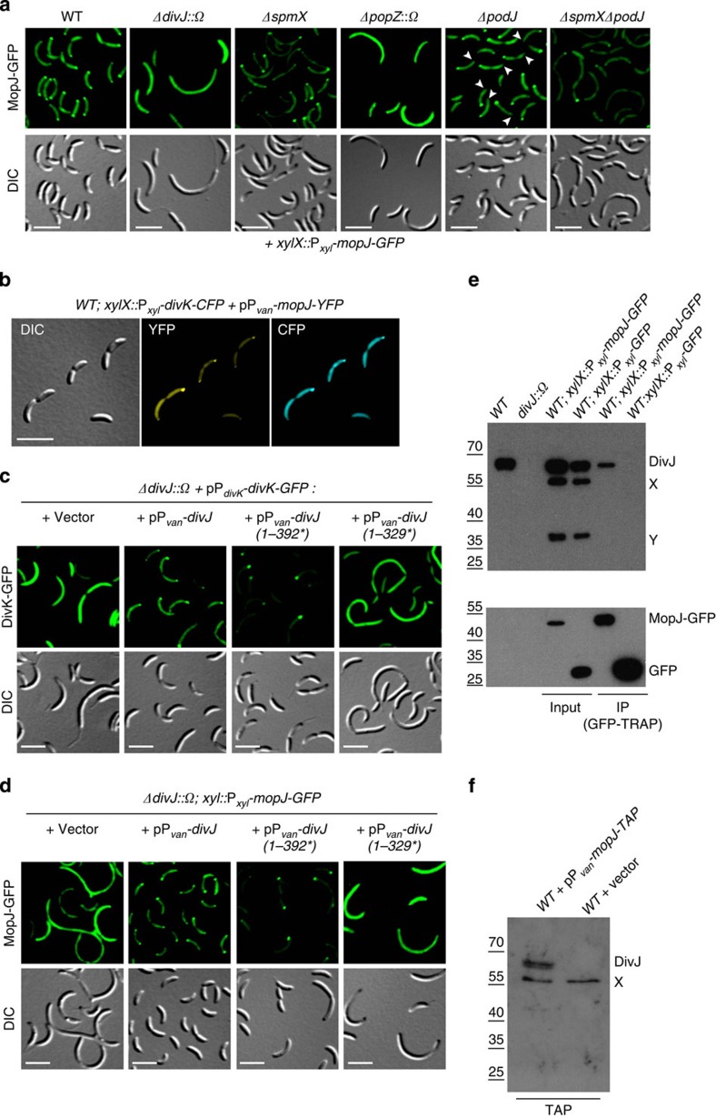
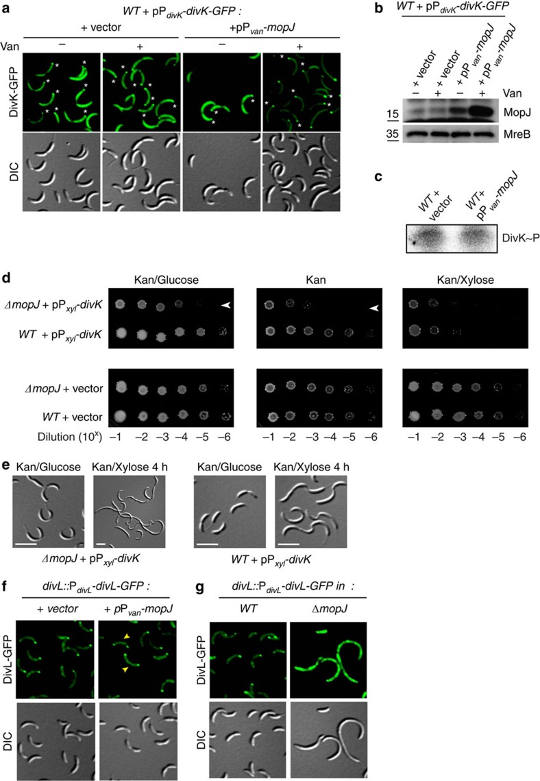
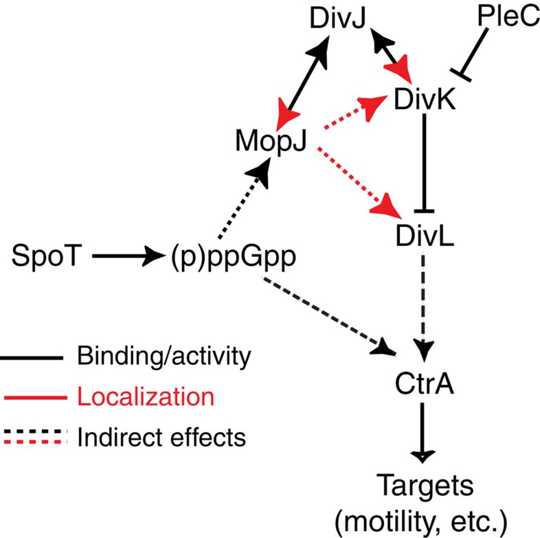
Despite the myriad of different sensory domains encoded in bacteria, only a few types are known to control the cell cycle. Here we use a forward genetic screen for Caulobacter crescentus motility mutants to identify a conserved single-domain PAS (Per-Arnt-Sim) protein (MopJ) with pleiotropic regulatory functions. MopJ promotes re-accumulation of the master cell cycle regulator CtrA after its proteolytic destruction is triggered by the DivJ kinase at the G1-S transition. MopJ and CtrA syntheses are coordinately induced in S-phase, followed by the sequestration of MopJ to cell poles in Caulobacter. Polarization requires Caulobacter DivJ and the PopZ polar organizer. MopJ interacts with DivJ and influences the localization and activity of downstream cell cycle effectors. Because MopJ abundance is upregulated in stationary phase and by the alarmone (p)ppGpp, conserved systemic signals acting on the cell cycle and growth phase control are genetically integrated through this conserved single PAS-domain protein.


Figures

Figure 1
Figure 1. MopJ, a pleiotropic regulator controlling motility and cell cycle progression in Caulobacter crescentus.
(a) Model showing the localization of His-Asp signalling proteins and its regulators along with MopJ during Caulobacter crescentus cell cycle. The black bars show the abundance of the transcriptional regulators CtrA (and phosphorylated CtrA, CtrA∼P) and GcrA. The grey arrow indicates when phosphorylation of the DivK receiver domain (RD) by the DivJ histidine kinase (HK) commences. Phosphorylated DivK (DivK∼P) is present henceforth. The thin vertical line in black represents the flagellum before it rotates (wavy line). The thick vertical line in black represents the stalk. The thin slanted black lines represent the polar pili. (b) (Upper panel) Motility assay on swarm agar plate of WT, mopJ::himar and ΔmopJ cells. (Lower panel) complementation of ΔmopJ cells harbouring the empty vector pMT335 or pMT335-mopJ. (c) Scheme of domain organization of MopJ from N- to C-terminus indicated by the total amino-acid (aa) length. The green arrowhead points to the residue in the mopJ coding sequence that was disrupted by the himar1 transposon insertion. (d,e) FACS analysis of WT and ΔmopJ cells. Genome content (FL1-A channel) and cell size (FSC-A channel) were analysed by FACS during exponential (d, Exp.) and stationary (e, Stat.) phases in M2G. G1 (1 N) and G2 cell (2 N) peaks of fluorescence intensity are readily visible (particularly in Stat. phase cells) and are labelled accordingly. S-phase cells are reflected by the broad intermittent signal (absent in Stat. phase cells). (f,g) Differential interference contrast (DIC) images of WT and ΔmopJ cells during Exp. (f) and Stat. (g) phases in M2G.
Figure 2
Figure 2. MopJ promotes CtrA accumulation and is regulated by (p)ppGpp.
(a,b) Promoter-probe assays of transcriptional reporters carrying the fljM, sciP, pilA or fljK promoter fused to a promoterless lacZ gene in WT and ΔmopJ cells in exponential (a, Exp.) and stationary (b, Stat.) phase. The graphs show lacZ-encoded β-galactosidase activities measured in Miller Units. (c) Immunoblot showing the steady-state levels of the major flagellin FljK, the SciP-negative regulator and the PilA structural subunit of the pilus filament in WT and ΔmopJ cells in Exp. and Stat. phase. The steady-state levels of the MreB actin are shown as a loading control (see Supplementary Fig. 4A). (d) Immunoblots showing CtrA and MopJ abundance in Exp. and Stat. phase ΔmopJ versus WT cells. Steady-state levels of MreB are shown as loading control (see Supplementary Fig. 4B). (e) Accumulation of CtrA is increased in ΔmopJ cells during Stat. phase either by inactivation of a proteolytic regulator encoded by cpdRcpdR::tet) or by appending an M2 (FLAG) epitope to the C-terminus of CtrA (ctrA::ctrA-M2). Immunoblot showing the steady-state levels of CtrA in WT, ΔmopJ, ΔcpdR::tet and ΔmopJ ΔcpdR::tet cells and the levels of CtrA-M2 in ctrA::ctrA-M2 and ΔmopJ ctrA::ctrA-M2 cells. Steady-state levels of MreB are shown as loading control. (f,g) Promoter-probe assays of transcriptional reporters carrying the PmopJ-lacZ in WT and ΔspoT cells in Exp. (f) and Stat. (g) phase. (h) LacZ assays with PmopJ-lacZ promoter-probe plasmid in WT and cells expressing the constitutive active form of E. coli RelA (RelA') and the catalytic mutant (RelA'-E335Q) fused to the FLAG tag in the presence of xylose. (a,b,f,g,h) Error bar (black bar) is shown as standard deviation (s.d., n=3).
Figure 3
Figure 3. Multi-layered spatiotemporal regulation of MopJ.
(a) Immunoblots showing steady-state levels of MopJ, GcrA, CtrA and DivJ in synchronized WT cells and MopJ in stationary (Stat.) phase (a, right, see Supplementary Fig. 5a,b). The red line marks the position of MopJ. (b) MopJ levels in exponential (Exp.) or stationary (Stat.) phase swarmer (SW) and stalked (ST)/predivisional (PD) cells detected using antibodies to MopJ (see Supplementary Fig. 5c). (c) LacZ measurements (Miller units) of PmopJ-lacZ in WT (blue) and two independent ΔgcrA xylX::Pxyl-gcrA (red and green) mutants, during 6 h (hours) of inhibition (glucose) of gcrA expression. Note the decline in activity upon dilution of overnight cells is due to the prior induction in stationary phase by (p)ppGpp (see Fig. 2f–h and Supplementary Fig. 3b). Error bar (black bar) is shown as s.d. (n=3). (d) Occupancy of GcrA (blue ovals) at the mopJ locus in WT cells as determined by chromatin immunoprecipitation coupled to deep-sequencing (ChIP-Seq) using polyclonal antibodies to GcrA. Occupancy is shown as percentage of total reads in the precipitated sample as a function of the chromosome coordinates. (e) Trace of N6-methyladenosine (m6A) marked DNA at mopJ locus in WT cells (blue trace) and ΔccrM::Ω (red trace) from ChIP-Seq experiment performed with antibodies to m6A. Occupancy is shown as percentage of total nucleotide coverage in sequencing as a function of the chromosome coordinates at the mopJ locus. Stars show the positions of 5′-GANTC-3′ sequences that are methylated by the CcrM methyltransferase at the N6 position of adenine. (fh) Immunoblots showing the steady-state levels of MopJ in ΔclpX xylX::Pxyl-clpX cells with (Xyl, xylose) or without (Glu, glucose) induction of Pxyl for 4 h (f), upon induction of the dominant negative form ClpX* from Pxyl for 4 h (g), and in WT, ΔsocB and ΔsocBΔclpX::Ω, ΔsocBΔclpP::Ω double mutants (h) along with CtrA and MreB (control). (i) Immunoblot showing MopJ-GFP and CtrA levels in synchronized mopJ::mopJ-GFP cells (see Supplementary Fig. 5d). (j,k) Time-lapse fluorescence imaging of live mopJ::mopJ-GFP cells (j) or WT xylX::Pxyl-mopJ-GFP cells (k). White asterisk denotes stalked pole. (a,i,j,k) Numbers indicate minutes after synchronization.
Figure 4
Figure 4. Polar localization of MopJ requires the DivJ kinase and the PopZ polar scaffold.
(a) Fluorescence and DIC images of live C. crescentus WT, ΔdivJ::Ω, ΔspmX, ΔpopZ::Ω, ΔpodJ or ΔpodJΔspmX cells expressing MopJ-GFP under the control of xylose promoter (Pxyl) at the xylX locus. White arrowheads point to the flagellated pole. (b) Co-localization of DivK-CFP expressed from Pxyl at the xylX locus in WT cells and MopJ-YFP expressed from Pvan on pMT374. The corresponding DIC image is also shown. (c) Fluorescence and DIC images of ΔdivJ::Ω cells expressing DivK-GFP fusion from pMR10-divK-GFP and various versions of DivJ, including full-length DivJ, DivJ-392 (short version of divJ encoding a truncated protein without histidine kinase domain) or DivJ-329 (short version of divJ encoding a truncated protein without kinase and dimerization domains) under the control of the vanillate promoter (Pvan) from pMT335. The control harbouring the empty vector is also shown. (d) Fluorescence and DIC images of ΔdivJ::Ω cells expressing MopJ-GFP under the control of Pxyl and the different forms of DivJ from pMT335 as described in c. (e) Co-immunoprecipitation (IP) of DivJ with MopJ-GFP from a GFP-TRAP affinity matrix (ChromoTek GmbH). Precipitated samples were probed for the presence of DivJ and GFP by immunoblotting using antibodies to DivJ (upper) and GFP (lower). Cell lysates used as input are also shown. (f) Tandem-affinity-purification (TAP) of MopJ-TAP from Pvan on pMT335 followed by immunoblotting of the samples for the presence of DivJ using polyclonal antibodies to DivJ. In e and f, X and Y denote nonspecific proteins that react with the antiserum or degradation products.
Figure 5
Figure 5. MopJ affects DivK localization and function.
(a) Fluorescence and DIC imaging of WT cells harbouring pMR10-divK-GFP and pMT335-Pvan-mopJ with (+) or without (−) induction (50 μM of vanillate) of MopJ from Pvan on pMT335. The controls harbouring pMR10-divK-GFP and the empty vector (pMT335) are also shown. The position of stalks are indicated by a white asterisk. (b) Immunoblot showing the steady-state levels of MopJ in strains used in a. Steady-state levels of MreB are shown as a loading control. (c) In vivo phosphorylation of cultures with 32P followed by immunoprecipitation reveals DivK∼P levels in WT cells harbouring pMT335 or pMT335-mopJ (Pvan-mopJ) after with vanillate (50 μM). (d) Effect of ΔmopJ mutation on viability in the presence of extra DivK. Efficiency of plating assays of WT and ΔmopJ cells harbouring the empty vector (pMR10) or the Pxyl-divK expression plasmid (pMR10-Pxyl-divK). Serial tenfold dilutions of cells plated on PYE agar with kanamycin and glucose (kan+gluc, glucose represses Pxyl), kanamycin (kan, background expression from Pxyl) and kanamycin and xylose (kan+xyl, xylose induces Pxyl) agar. (e) DIC (differential interference contrast) imaging of cells described in d in glucose or 4 h after induction with xylose (0.3%). The medium was supplemented with kanamycin (5 μg ml−1). (f) Effect of MopJ overexpression on DivL-GFP localization. The empty vector (pMT335) or the Pvan-mopJ plasmid (pMT335-mopJ) was transformed into divL::PdivL-divL-GFP cells and the resulting cells imaged by DIC and fluorescence microscopy 4 h after induction with vanillate (50 μM). Yellow arrowheads point to cells with bipolar DivL-GFP. (g) Effect of mopJ deletion on DivL-GFP localization. The divL::PdivL-divL-GFP allele was transduced in WT and ΔmopJ cells and the resulting cells imaged by DIC and fluorescence microscopy.
Figure 6
Figure 6. Proposed action of MopJ on the Caulobacter cell circuitry.
Summary scheme depicting the interactions of MopJ with the cell circuitry as determined in this work. Effects on protein localization are shown in red, whereas interactions shown in black designate direct interactions that generally result in activation (arrow, DivJ-DivK) or inhibition (T-bar, PleC-DivK, DivK-DivL). The scheme also shows the effects of the alarmone (p)ppGpp in inducing MopJ and the maintenance of CtrA (see Discussion).

Similar articles

Cited by

References

    1. Guttenplan S. B. & Kearns D. B. Regulation of flagellar motility during biofilm formation. FEMS Microbiol. Rev. 37, 849–871 (2013). - PMC - PubMed
    1. Skerker J. M. & Laub M. T. Cell-cycle progression and the generation of asymmetry in Caulobacter crescentus. Nat. Rev. Microbiol. 2, 325–337 (2004). - PubMed
    1. Radhakrishnan S. K., Thanbichler M. & Viollier P. H. The dynamic interplay between a cell fate determinant and a lysozyme homolog drives the asymmetric division cycle of Caulobacter crescentus. Genes Dev. 22, 212–225 (2008). - PMC - PubMed
    1. Quon K. C., Marczynski G. T. & Shapiro L. Cell cycle control by an essential bacterial two-component signal transduction protein. Cell 84, 83–93 (1996). - PubMed
    1. Ely B. Genetics of Caulobacter crescentus. Methods Enzymol. 204, 372–384 (1991). - PubMed

Publication types

Substances

LinkOut - more resources