. 2015 May 8:6:7056.
doi: 10.1038/ncomms8056.

Clk post-transcriptional control denoises circadian transcription both temporally and spatially

Affiliations

Clk post-transcriptional control denoises circadian transcription both temporally and spatially

Immanuel Lerner et al. Nat Commun. .

Abstract

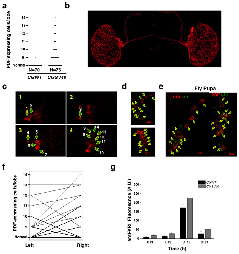
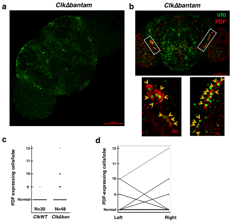
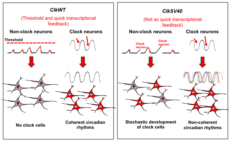
The transcription factor CLOCK (CLK) is essential for the development and maintenance of circadian rhythms in Drosophila. However, little is known about how CLK levels are controlled. Here we show that Clk mRNA is strongly regulated post-transcriptionally through its 3' UTR. Flies expressing Clk transgenes without normal 3' UTR exhibit variable CLK-driven transcription and circadian behaviour as well as ectopic expression of CLK-target genes in the brain. In these flies, the number of the key circadian neurons differs stochastically between individuals and within the two hemispheres of the same brain. Moreover, flies carrying Clk transgenes with deletions in the binding sites for the miRNA bantam have stochastic number of pacemaker neurons, suggesting that this miRNA mediates the deterministic expression of CLK. Overall our results demonstrate a key role of Clk post-transcriptional control in stabilizing circadian transcription, which is essential for proper development and maintenance of circadian rhythms in Drosophila.


Conflict of interest statement

Competing Financial Interest Statement: The authors declare that there is no conflict of interest.

Figures

Figure 1
Figure 1. Clk is under strong post-transcriptional regulation
a. Comparison of clock transcript levels. RNA-seq data was used to compare expression levels of core circadian components in fly heads; an average of six timepoints ± SE in LD conditions (ZT 2,6,10,14,18,22) were analyzed. b. The levels of pre-mRNA and mRNA of core clock components were measured by real-time PCR (RT-PCR) from a mixture of six circadian time points and scaled using an equimolar mixture of the PCR products to obtain absolute measurements between the different mRNAs or pre-mRNAs in each sample (mean ± SE; three biological replicas). As the values in each sample (pre-mRNA and mRNA) are normalized, the pre-mRNA/mRNA ratio is relative, not absolute (see methods for details). c. RT-PCR measurements of transcriptional (gray) and steady state (black) levels of Clk and tim. Transcriptional/pre mRNA transcripts were detected using primers for intronic sequences of the genes, whereas steady state/ mature mRNAs were detected using primers flanking two exons. Expression was normalized to RP49 and RpS18. Representative experiment of three repeats is shown. d. RT-PCR results presenting the ratio of mRNA bound to oligo-dT beads vs. unbound mRNA. Data pooled from a mixture of six time points (mean ± SE; three biological replicates). e. A Drosophila fly wing system was developed to monitor circadian gene mRNA expression levels (normalized to RP49 at ZT5, 17). Clk and tim levels cycle with the expected phase. The measurements were performed in triplicated for each time point using the Canton-S strain. f. RT-PCR mRNA half-life measurements for Clk and cry mRNAs from fly wings treated with actinomycin D in five different time time-points (0-4h after actinomycin D exposure, 12 h light/12 hour dark conditions,mean ± SE; three biological replicates). See also Supplementary Figure 1
Figure 2
Figure 2. Clk 3’ UTR establishes a threshold for CLK-driven transcription
a. Schematics of the system for monitoring post-transcriptional regulation in single Drosophila S2 cells. b. Mean fluorescent levels of S2 cell population expressing the Clk and SV40 3’ UTR fluorescent reporters at different copper levels. The inset in the left represents a closer view of the signal at low copper concentrations (0-15 µM). Cyan fluorescent protein (CFP) expressed from a pAc-CFP plasmid, was utilized as transfection control. c. Schematics of the ClkSV40 and tim-YFP constructs. d. Histogram representing the intensity of signal due to the tim-YFP reporter upon transfection with ClkSV40 or ClkWT plasmids. See also Supplementary Figure 2. Red fluorescent protein (RFP) expressed from a pAc-RFP plasmid, was utilized as transfection control.
Figure 3
Figure 3. ClkSV40 flies express a stable mRNA that generates ectopic clock cells
a. AGO1 binding to endogenous genomic Clk mRNA or ClkV5 from ClkWT or ClkSV40 transgenic fly heads (measured by RT-PCR, ratio of IP vs input is shown, one representative experiment out of three is presented). b. RT-PCR results showing the ratio of Clk mRNA bound vs. unbound- to oligo-dt beads in ClkWT and ClkSV40 fly heads (* p<0.036, student t-test; mean ± SE, n=6 for each genotype, average of five independent insertions of these transgenes were plotted). c, d. Immunofluorescence (IF) analysis of (c) ClkSV40 or (d) ClkWT Drosophila brains using an anti-V5 antibody (lower panel represents magnification of rectangle area). e. Expression levels of CLKV5, total CLK, and tubulin proteins determined by western blot analysis of head lysates from ClkWT or ClkSV40 flies (five independent insertions for each transgene). The showed experiment is one out of four representative experiments. f, g. Immunofluorescence analysis of (f) ClkSV40 or (g) ClkWT Drosophila brains using an anti-VRI antibody. h, i. ClkV5 and endogenous Clk mRNAs levels from ClkSV40 and ClkWT fly heads determined by RT-PCR from (h) total RNA and (i) polyA+-selected RNA (for each time point, the average of five independent insertions for each transgene is plotted). See also Supplementary Movie 1 and Supplementary Movie 2.
Figure 4
Figure 4. ClkSV40 flies display variable circadian behavior
a. Bar graph summarizing the behavioral analyses of ClkWT and ClkSV40 flies. Each row and set of numbers (e.g. 1-1) represents a strain with an independent insertion of the wild type (ClkWT) or SV40 (ClkSV40) transgene. In all cases flies carry one extra copy of the Clk gene (either ClkWT or ClkSV40) in a wild-type background. Flies were classified as rhythmic or arrhythmic in the first 5 days in constant darkness using the Behavioral toolbox . b. Classification of rhythmic flies as normal and atypical. The classification was done manually after 10 days in constant darkness. c. Examples of individual ClkSV40 flies displaying atypical circadian behavior. See also Supplementary Figure 3 and 4.
Figure 5
Figure 5. Stochastic development of the circadian system in ClkSV40 flies
a. Number of PDF-positive cell bodies in ClkWT and ClkSV40 brains. Data is from the ClkSV40 (2-8) and ClkWT (1-1) fly strains. b. Representative example of a ClkSV40 fly brain in which the position of extra pdf-expressing cells can be visualized. c. Representative example of a ClkSV40 fly brain in which 14 pdf-expressing cells were observed in one of the brain hemispheres. Z stack of 3 images (each taken every 1 µm). Numbers in the upper left corner of each picture indicate the relative position of the Z stack. Arrows indicate the position of the pdf-expressing cells. d. Z stack (maximal projection) of 2 brain hemispheres of ClkSV40 flies immunostained for PDF showing the number of pdf-expressing cells (13 and 12 for the top and bottom brain hemispheres respectively). Arrows indicate the position of the pdf-expressing cells. Red scale bar indicates 10μm. e. Z stack (maximal projection) of 2 brains of ClkSV40 pupae immunostained for PDF (in red) and VRI (in green) showing the number of pdf-expressing cells (14 and 15 for the left and right images respectively). Arrows indicate the position of the pdf-expressing cells. Red scale bar indicates 10μm. f. Comparison of left and right hemispheres in ClkWT and ClkSV40 flies based on PDF immunofluorescent staining. g. Brain to brain variation in the levels of VRI (measured by IF) protein in the sLNvs at 4 time points (day 10 in DD), in ClkSV40 (line 2-8, red) and ClkWT (line 1-1, black) flies. See also Supplementary Figure 5, 6, 7 and 8 Supplementary Movies 3 and 4.
Figure 6
Figure 6. Deletion of the bantam binding sites on the Clk 3’UTR lead to stochastic development of the pdf-expressing cells
a. Immunofluorescence (IF) analysis of a representative ClkΔban (3-7) Drosophila brain using an anti-VRI antibody. Flies were collected and dissected at ZT15. b. Representative example of a ClkΔban fly brain in which 21 LNvs cells were observed, 9 in one hemisphere and 12 in the other (top panel). Lower panel represents magnification of rectangle area. Of the 12 PDF positive cell bodies in the right hemisphere: 9 cells are VRI positive, one cell display low intensity of VRI immunostaining and 2 cells are VRI negative. In the left hemisphere of the brain, 9 PDF positive cells are observed: 8 cells are VRI positive and one cell displays low intensity of VRI immunostaining. Flies were collected and dissected at ZT15. Arrows indicate the position of each PDF positive cell. c. Number of PDF-positive cell bodies in ClkWT and ClkΔban brains. Data is taken from the ClkΔban (3-7) and ClkWT (1-1) fly strains. d. Comparison of left and right hemispheres in ClkWT and ClkΔban flies.
Figure 7
Figure 7. Role of Clk post-transcriptional regulation in clock and non-clock neurons
Small red arrows represent random or environmentally driven fluctuations in Clk gene expression.

Similar articles

Cited by

References

    1. Allada R, Chung BY. Circadian organization of behavior and physiology in Drosophila. Annu Rev Physiol. 2010;72:605–24. - PMC - PubMed
    1. Hall JC. Genetics and molecular biology of rhythms in Drosophila and other insects. Adv Genet. 2003;48:1–280. - PubMed
    1. Allada R, White NE, So WV, Hall JC, Rosbash M. A mutant Drosophila homolog of mammalian Clock disrupts circadian rhythms and transcription of period and timeless. Cell. 1998;93:791–804. - PubMed
    1. Rutila JE, et al. CYCLE is a second bHLH-PAS clock protein essential for circadian rhythmicity and transcription of Drosophila period and timeless. Cell. 1998;93:805–14. - PubMed
    1. Hardin PE, Hall JC, Rosbash M. Feedback of the Drosophila period gene product on circadian cycling of its messenger RNA levels. Nature. 1990;343:536–40. - PubMed

Publication types

MeSH terms

LinkOut - more resources